- Home
- Staff Profiles & Phone Book
- About the Department
- Study Anatomy
- Study Neuroscience
- Research
- UCC Anatomical Donations
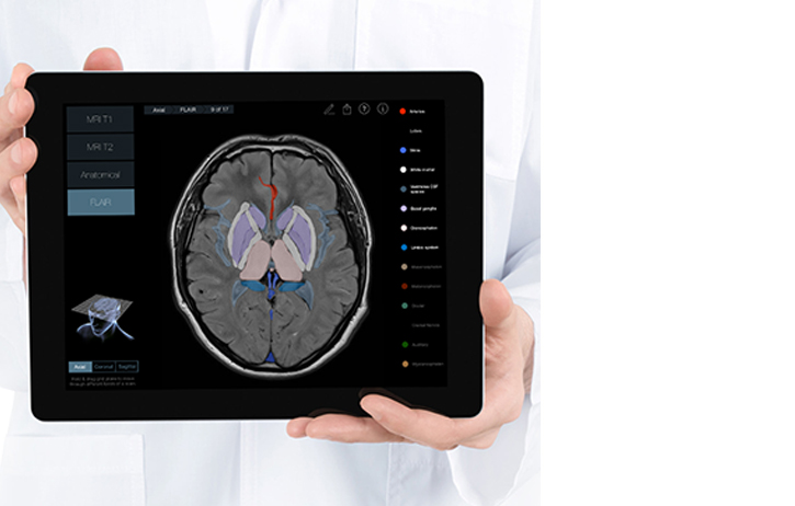
- Biosciences Imaging Centre
- BSc Medical and Health Sciences
- News & Events
- BRAIN AWARENESS WEEK 2023
- NEWS ARCHIVE 2023
- News Archive 2022
- News Archive 2021
- News Archive 2020
- News Archive 2019
- News Archive 2018
- News archive 2017
- News Archive 2016
- News Archive2015
- News Archive 2014
- News Archive 2013
- News Archive 2012
- News Archive 2011
- Department Events and Conferences
- Seminar series 2019_2020
- Recent Publications
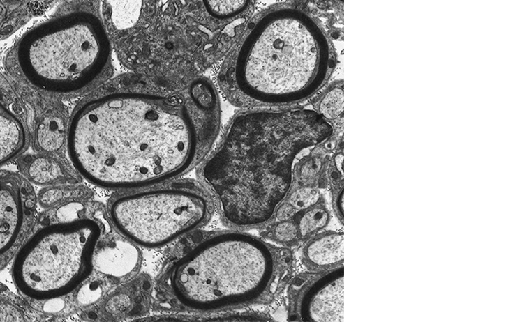
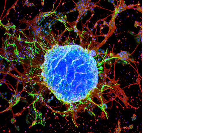
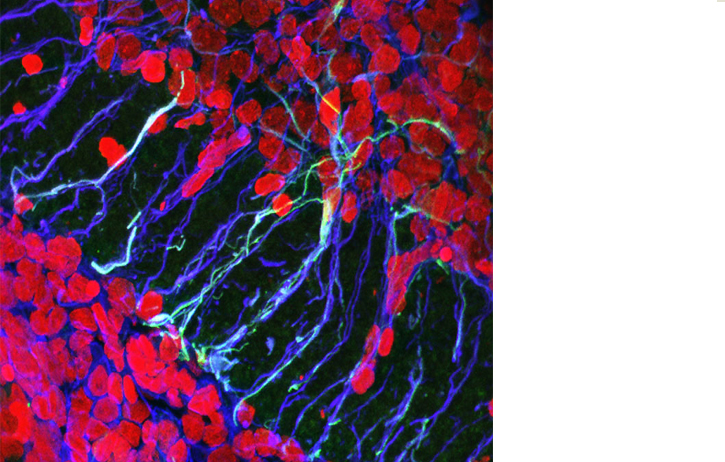
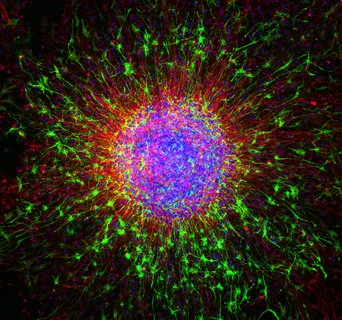
- photo galleries
- Contact Us
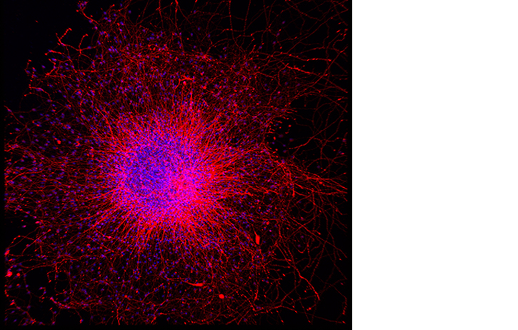
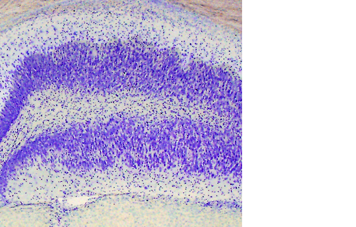
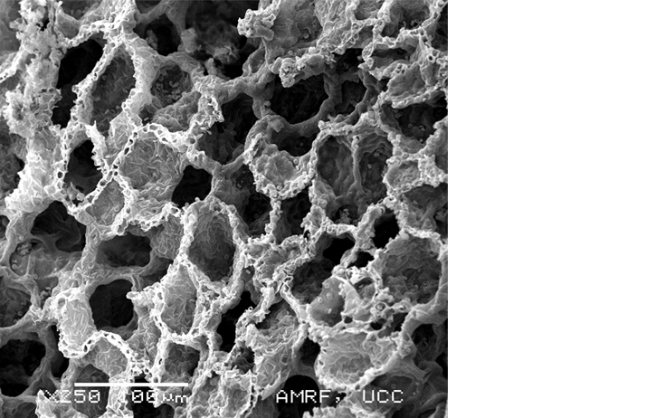
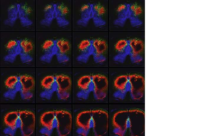
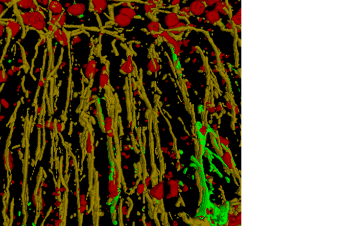
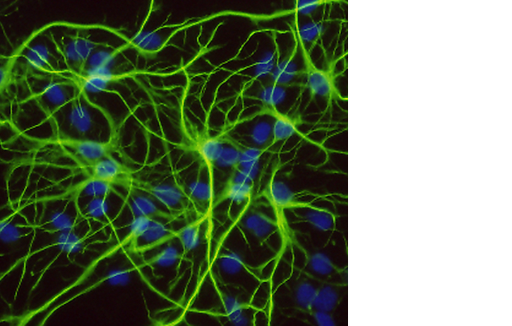
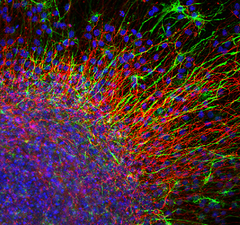
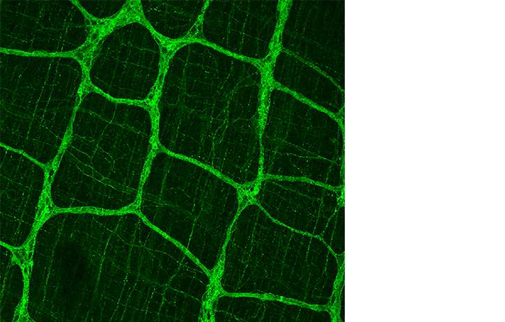
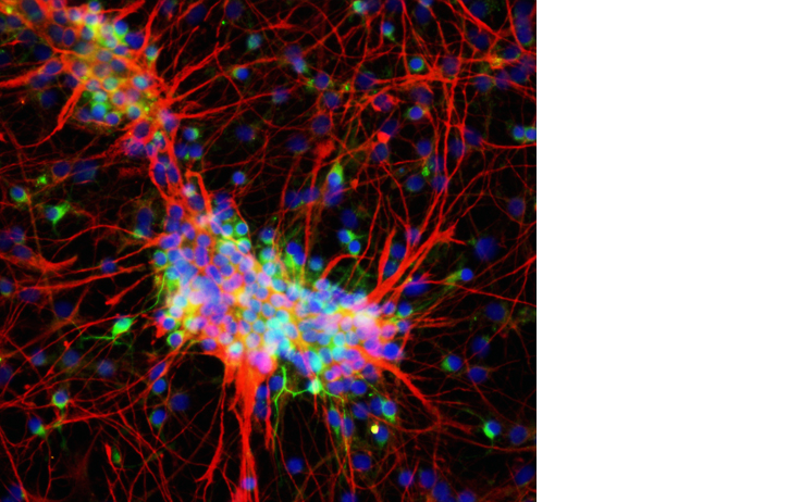
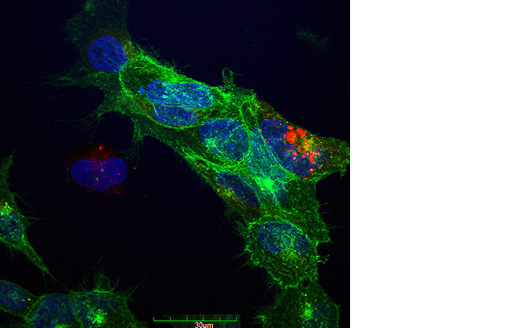
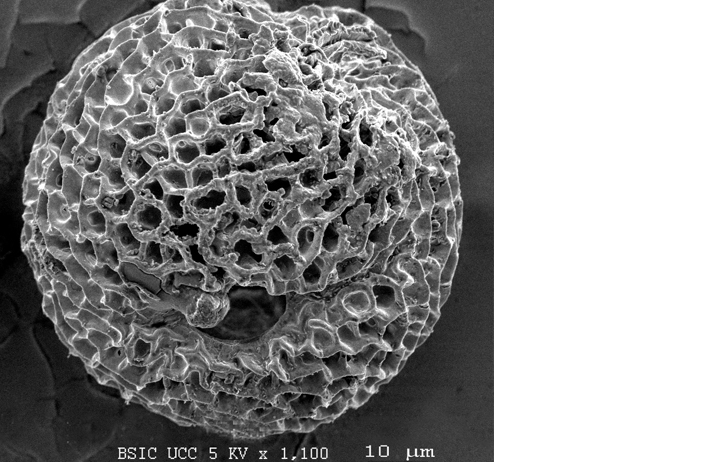
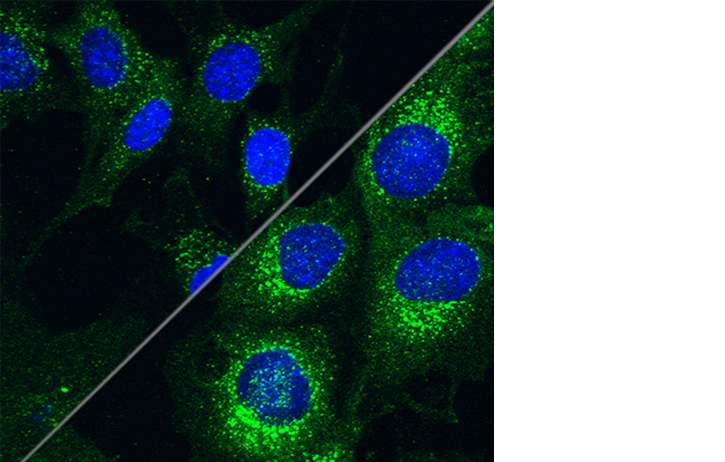
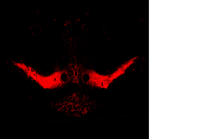
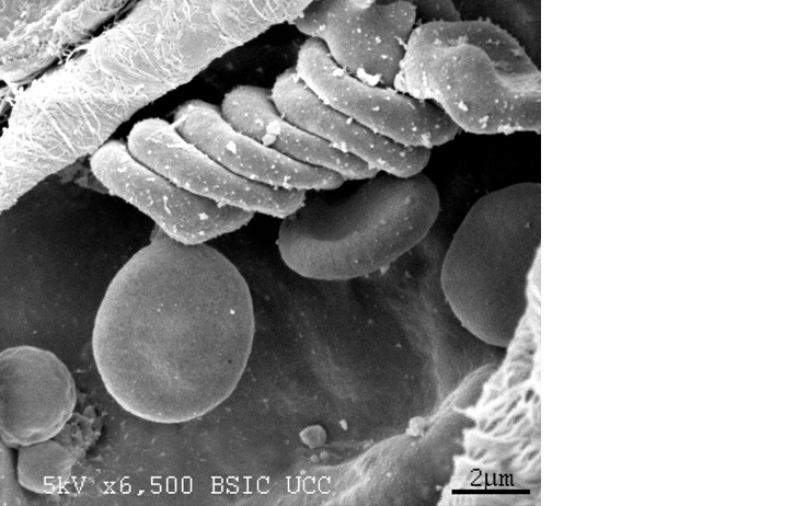
IMAGE OF THE MONTH
Archive of Image of the Month
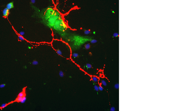
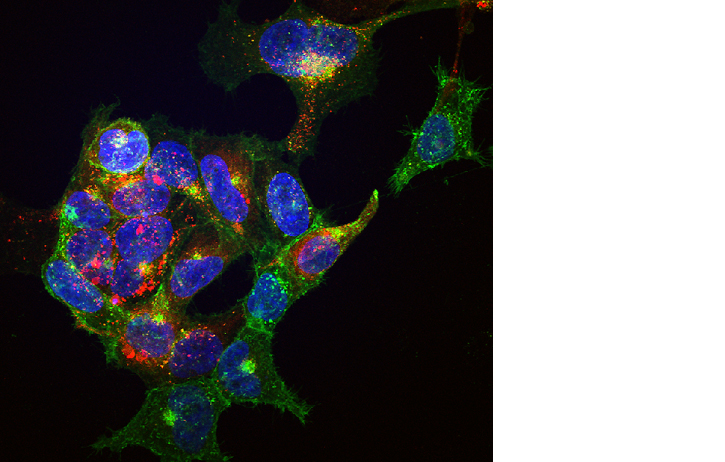
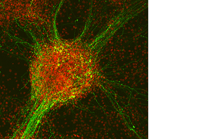
Click on the images below to scroll through and have a closer look at some of our previous images of the month. Click on READ MORE tab above to read details about all images of the month.
,SOX2(blue),July2014.jpg)

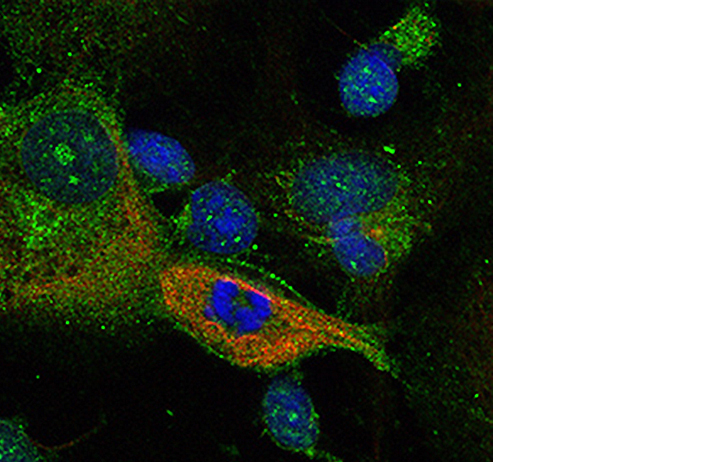
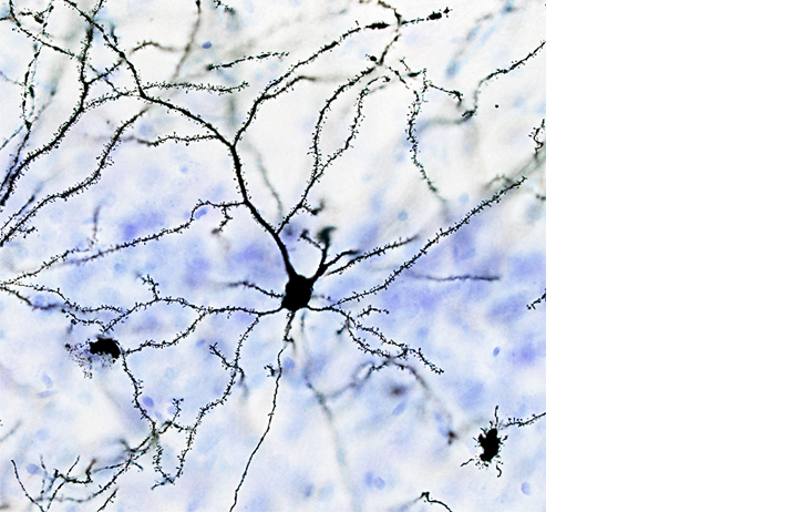
February
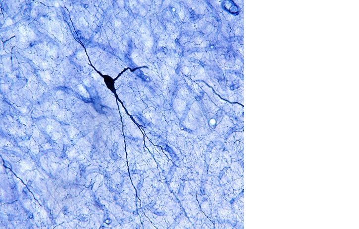
February 2016: "Expression of NADPH diaphorase (a marker of nitric oxide synthase (NOS) enzyme activity) in rat cortical neurons"
Submitted by: Submitted by Dr Eoin Sherwin
Dr Eoin Sherwin is a post-doctoral researcher with the APC Microbiome institute. His research focuses on the development of live biotherapeutics for the treatment of autistic spectrum disorders.
The image depicts rat cortical neurons that has been stained with NADPH diaphorase. NADPH diaporase staining is indicative of cells that are actively producing the free radical gas, nitric oxide, via the enzymatic actions of nitric oxide synthase."