In This Section
- Home
- Staff Profiles & Phone Book
- About the Department
- Welcome from Head of Department of Anatomy and Neuroscience
- A History of the Department
- A history of the Department; The early years to the 1980s
- A history of the Department; The move from the Windle Building to BSI and WGB
- UCC Professors of Anatomy and Heads of Department
- The development of the UCC HUB
- Current students, recent research graduates and awards
- Useful Links
- Study Anatomy
- Study Neuroscience
- Research
- Neural circuitry underlying Neuropsychiatric and Neurological Disorders 2026
- Neurogastroenterology 2026
- Developmental Neuroscience and Regeneration 2026
- Neurodegeneration 2026
- Neuroinflammation 2026
- Neuroprotection and Therapeutics 2026
- Neuroproteomics and Molecular Psychiatry 2026
- Anatomy Education Research 2026
- Research Facilities 2026
- Postgraduate Research Programmes 2026
- UCC Anatomical Donations
- Biosciences Imaging Centre
- BSc Medical and Health Sciences
- News & Events
- News Archive 2024
- News Archive 2023
- News Archive 2022
- News Archive 2021
- News Archive 2020
- News Archive 2019
- News Archive 2018
- News archive 2017
- News Archive 2016
- News Archive2015
- News Archive 2014
- News Archive 2013
- News Archive 2012
- News Archive 2011
- BRAIN AWARENESS WEEK 2023
- Department Events and Conferences
- Seminar series 2019_2020
- photo galleries
- Narrowing the void Conference 2023
- Photos of BSc Medical and Health Sciences Mentoring launch 2022
- International Women's Day 2023
- 2023 BRIGHT FUTURES - Celebrating our researchers
- 2023 UCC Futures - Future Ageing & Brain Sciences
- Recent Graduations July 2023
- Anatomy and Neuroscience Top 100 Anatomy Physiology 2023
- BRAIN AWARENESS WEEK 2023 FUN AND GAMES EVENT
- Medical and Health Sciences First year class 2023
- 2023 Brain Awareness week Scientific discussion photo gallery
- World Anatomy Day 2023
- BSc MHS MENTORING PROGRAMME 2023
- BSc Medical and Health Sciences Graduation 2023
- BSc Neuroscience Graduation Photo Gallery 2023
- Dr Kathy Quane Nov 2023
- THANKSGIVING PHOTOS 2012
- Photo Gallery: Society of Translational Medicine Careers Fair 2023
- Photo Gallery:2023 TRAIN AWARDS
- Photo Gallery:2024 Creative Week St Joseph's NS
- Photo Gallery: Department of Anatomy and Neuroscience Thanksgiving Service 2024
- Photo Gallery: Professor Aideen Sullivan farewell party
- Photo Gallery: Irish Pain Society Annual Scientific Meeting Cork 2023
- Photo Gallery: 2024 Medical and Health Sciences Graduation
- Photo Gallery: Medical and Health Sciences Meet and Greet 2024
- Photo Gallery: 2024 BSC NEUROSCIENCE Graduation
- Photo Gallery: 2025 INTERNATIONAL WOMEN'S DAY
- Photo Gallery: 2025 BSc Neuroscience class and staff
- Photo Gallery: 2025 BRAIN CONNECTIONS
- BSc Neuroscience Graduation Photo Gallery 2025
- World Anatomy Day 2025
- UCC Learning and Teaching Showcase 2025
- MSc Human Anatomy Graduation Photo Gallery 2025
- Narrowing the Void Conference 2023
- Department of Anatomy and Neuroscience Contact Us
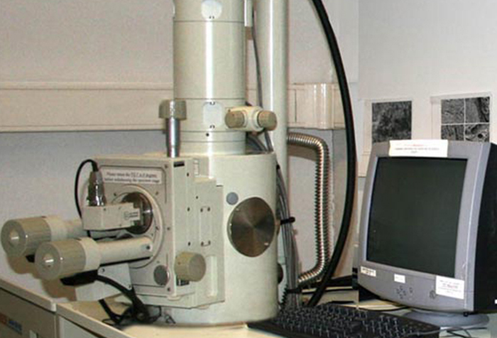
Joel JSM 5510 SEM

Jeol Scanning Electron Microscope JSM-5510
Jeol Scanning Electron Microscope JSM-5510 (Jeol Ltd., Tokyo, Japan) is available forScanning Electron Microscopy.
The Jeol Scanning Electron Microscope (JSM 5510) operates at voltages up to 30kV, and has an Oxford Instruments Energy Dispersive X-Ray Spectroscopy detector attachment for elemental analysis.
The scanning electron microscope (SEM) images the sample surface by scanning it with a high-energy beam of electrons in a raster scan pattern. The beam of electrons strikes the surface of the specimen and interacts with the sample at or near its surface, producing signals that contain information about the sample. The types of signals generated by an SEM include secondary electrons, back scattered electrons, characteristic x-rays and light. Electronic devices are used to detect and amplify the secondary electrons and display them as an image which is digitally captured and displayed on a computer monitor. The SEM can produce very high-resolution images of a sample surface, revealing details less than 5 nm in size. SEM micrographs have a very large depth of focus yielding a characteristic three-dimensional appearance useful for understanding the surface structure of a sample.
The SEM in the EM Facility is currently used to study samples from a variety of projects, from many different disciplines, including materials science, food science, biological sciences and geology. In all cases specimens must be dry, conductive and able to withstand the high vacuum present inside the instrument.
Composite image showing left to right: Jeol Scanning Electron Microscope (JSM 5510), Beetle maxilla x900, pollen grain x3000, rat trachea x2500, rat tongue x400 ,pollen grains, silicon chip x 450. Images S Crotty
Department of Anatomy and Neuroscience
Anatamaíocht agus Néareolaíocht
Contact us
Room 2.33, 2nd Floor, Western Gateway Building, University College, Cork, Ireland
