In This Section
- Home
- About the Department
- Welcome from Head of Department of Anatomy and Neuroscience
- A History of the Department
- A history of the Department; The early years to the 1980s
- A history of the Department; The move from the Windle Building to BSI and WGB
- UCC Professors of Anatomy and Heads of Department
- The development of the UCC HUB
- Current students, recent research graduates and awards
- Useful Links
- People and Phonebook
- Study Anatomy
- Study Neuroscience
- Research
- Neural circuitry underlying Neuropsychiatric and Neurological Disorders 2026
- Neurogastroenterology 2026
- Developmental Neuroscience and Regeneration 2026
- Neurodegeneration 2026
- Neuroinflammation 2026
- Neuroprotection and Therapeutics 2026
- Neuroproteomics and Molecular Psychiatry 2026
- Anatomy Education Research 2026
- Research Facilities 2026
- Postgraduate Research Programmes 2026
- UCC Anatomical Donations
- Biosciences Imaging Centre
- News & Events
- 2025 News Archive
- News Archive 2024
- News Archive 2023
- News Archive 2022
- News Archive 2021
- News Archive 2020
- News Archive 2019
- News Archive 2018
- News archive 2017
- News Archive 2016
- News Archive2015
- News Archive 2014
- News Archive 2013
- News Archive 2012
- News Archive 2011
- BRAIN AWARENESS WEEK 2023
- Department Events and Conferences
- Seminar series 2019_2020
- photo galleries
- Narrowing the void Conference 2023
- Photos of BSc Medical and Health Sciences Mentoring launch 2022
- International Women's Day 2023
- 2023 BRIGHT FUTURES - Celebrating our researchers
- 2023 UCC Futures - Future Ageing & Brain Sciences
- Recent Graduations July 2023
- Anatomy and Neuroscience Top 100 Anatomy Physiology 2023
- BRAIN AWARENESS WEEK 2023 FUN AND GAMES EVENT
- Medical and Health Sciences First year class 2023
- 2023 Brain Awareness week Scientific discussion photo gallery
- World Anatomy Day 2023
- BSc MHS MENTORING PROGRAMME 2023
- BSc Medical and Health Sciences Graduation 2023
- BSc Neuroscience Graduation Photo Gallery 2023
- Dr Kathy Quane Nov 2023
- THANKSGIVING PHOTOS 2012
- Photo Gallery: Society of Translational Medicine Careers Fair 2023
- Photo Gallery:2023 TRAIN AWARDS
- Photo Gallery:2024 Creative Week St Joseph's NS
- Photo Gallery: Department of Anatomy and Neuroscience Thanksgiving Service 2024
- Photo Gallery: Professor Aideen Sullivan farewell party
- Photo Gallery: Irish Pain Society Annual Scientific Meeting Cork 2023
- Photo Gallery: 2024 Medical and Health Sciences Graduation
- Photo Gallery: Medical and Health Sciences Meet and Greet 2024
- Photo Gallery: 2024 BSC NEUROSCIENCE Graduation
- Photo Gallery: 2025 INTERNATIONAL WOMEN'S DAY
- Photo Gallery: 2025 BSc Neuroscience class and staff
- Photo Gallery: 2025 BRAIN CONNECTIONS
- BSc Neuroscience Graduation Photo Gallery 2025
- World Anatomy Day 2025
- UCC Learning and Teaching Showcase 2025
- MSc Human Anatomy Graduation Photo Gallery 2025
- Department of Anatomy & Neuroscience DELTA AWARD 2026
- Thanksgiving Service 2026
- BSc Neuroscience Class photos 2026
- Flame Sculpture by Alexandra Wejchert
- FLAME LABORATORY GALLERY
- Narrowing the Void Conference 2023
- Department of Anatomy and Neuroscience Contact Us
Lecturers in the Department of Anatomy & Neuroscience deliver innovative 3D dental anatomy teaching and examination in Canvas
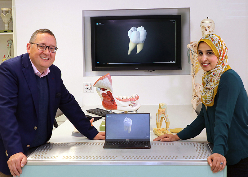
Dr André Toulouse and Dr Mutahira Lone, lecturers in the Department of Anatomy and Neuroscience, have developed an innovative blended learning approach which incorporates online 3D models for teaching and examining of dental morphology into Canvas.
During the past academic year, the implementation of COVID-19-related restrictions limited student attendance on campus. This introduced issues for the delivery of the AN2008 - The Dental Morphology, Histology and Embryology module, as one of the main learning outcomes is the capacity of learners to recognise the three-dimensional features of teeth and identify individual teeth. The module is usually taught using extracted teeth and resin models.
To circumvent some of the issues, Drs André Toulouse and Mutahira Lone, from the Department of Anatomy and Neuroscience, developed an innovative blended learning approach that included online 3D models for teaching and examining of dental morphology. The teaching material made available to students included a number of digital teaching tools such as lecture recordings, lecture notes, 2D dental morphology cue cards, but in particular 3D models of individual teeth were embedded in the Canvas VLE platform for study. The models could be accessed through an interactive dental charting grid, could be rotated freely by the learner and had key identification features labelled. Unlabelled models were also used during live interactive sessions with the lecturers and practice quizzes.
The examination was a Canvas VLE quiz where the students had limited time to identify previously unseen 3D models of a teeth and give the correct answer using a dental notation system. Results were in line with previous examinations performed using extracted teeth, suggesting that students have achieved some of the learning objectives. Feedback collected shows that the students used these 3D models extensively for studying tooth morphology and that models were their preferred learning tools together with the live interactive sessions. The results of this study are published in Journal of Dental Education.
This is the first time that the Canvas VLE was used for 3D teaching and examination in the Department of Anatomy and Neuroscience.
Photograph B.Riedewald
Department of Anatomy and Neuroscience
Anatamaíocht agus Néareolaíocht
Contact us
Room 2.33, 2nd Floor, Western Gateway Building, University College, Cork, Ireland