In This Section
- Home
- Staff Profiles & Phone Book
- About the Department
- Welcome from Head of Department of Anatomy and Neuroscience
- A History of the Department
- A history of the Department; The early years to the 1980s
- A history of the Department; The move from the Windle Building to BSI and WGB
- UCC Professors of Anatomy and Heads of Department
- The development of the UCC HUB
- Current students, recent research graduates and awards
- Useful Links
- Study Anatomy
- Study Neuroscience
- Research
- Neural circuitry underlying Neuropsychiatric and Neurological Disorders 2026
- Neurogastroenterology 2026
- Developmental Neuroscience and Regeneration 2026
- Neurodegeneration 2026
- Neuroinflammation 2026
- Neuroprotection and Therapeutics 2026
- Neuroproteomics and Molecular Psychiatry 2026
- Anatomy Education Research 2026
- Research Facilities 2026
- Postgraduate Research Programmes 2026
- UCC Anatomical Donations
- Biosciences Imaging Centre
- BSc Medical and Health Sciences
- News & Events
- News Archive 2024
- News Archive 2023
- News Archive 2022
- News Archive 2021
- News Archive 2020
- News Archive 2019
- News Archive 2018
- News archive 2017
- News Archive 2016
- News Archive2015
- News Archive 2014
- News Archive 2013
- News Archive 2012
- News Archive 2011
- BRAIN AWARENESS WEEK 2023
- Department Events and Conferences
- Seminar series 2019_2020
- photo galleries
- Narrowing the void Conference 2023
- Photos of BSc Medical and Health Sciences Mentoring launch 2022
- International Women's Day 2023
- 2023 BRIGHT FUTURES - Celebrating our researchers
- 2023 UCC Futures - Future Ageing & Brain Sciences
- Recent Graduations July 2023
- Anatomy and Neuroscience Top 100 Anatomy Physiology 2023
- BRAIN AWARENESS WEEK 2023 FUN AND GAMES EVENT
- Medical and Health Sciences First year class 2023
- 2023 Brain Awareness week Scientific discussion photo gallery
- World Anatomy Day 2023
- BSc MHS MENTORING PROGRAMME 2023
- BSc Medical and Health Sciences Graduation 2023
- BSc Neuroscience Graduation Photo Gallery 2023
- Dr Kathy Quane Nov 2023
- THANKSGIVING PHOTOS 2012
- Photo Gallery: Society of Translational Medicine Careers Fair 2023
- Photo Gallery:2023 TRAIN AWARDS
- Photo Gallery:2024 Creative Week St Joseph's NS
- Photo Gallery: Department of Anatomy and Neuroscience Thanksgiving Service 2024
- Photo Gallery: Professor Aideen Sullivan farewell party
- Photo Gallery: Irish Pain Society Annual Scientific Meeting Cork 2023
- Photo Gallery: 2024 Medical and Health Sciences Graduation
- Photo Gallery: Medical and Health Sciences Meet and Greet 2024
- Photo Gallery: 2024 BSC NEUROSCIENCE Graduation
- Photo Gallery: 2025 INTERNATIONAL WOMEN'S DAY
- Photo Gallery: 2025 BSc Neuroscience class and staff
- Photo Gallery: 2025 BRAIN CONNECTIONS
- BSc Neuroscience Graduation Photo Gallery 2025
- World Anatomy Day 2025
- UCC Learning and Teaching Showcase 2025
- MSc Human Anatomy Graduation Photo Gallery 2025
- Narrowing the Void Conference 2023
- Department of Anatomy and Neuroscience Contact Us
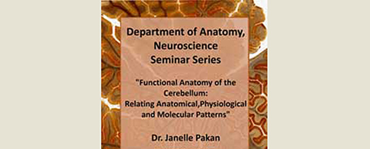
Anatomy Department Neuroscience Seminar Series

Dr. Janelle Pakan is a Post-Doctoral Fellow in the Department of Anatomyat UCC. She received her PhD in Neuroscience from the University of Alberta, Canada where she studied cerebellar organization in relation tovisual processing and sparked her interest in microscopy and imaging techniques.
Janelle then spent a year in Vancouver, Canada as a Post-Doctoral Fellow where her focus turned to functional properties ofneuroglia and imaging techniques associated with observing glial behaviourin living tissue.
She is now working with Dr. Kieran McDermott at UCC,using multiphoton imaging to study cell migration and differentiation in thedeveloping spinal cord.
Abstract
Cerebellum is Latin for 'little brain', however the cerebellum contains over halfof the total number of neurons in our brain. If it follows that size matters, it isobviously a very important structure. Traditionally, the cerebellum was thoughtto be involved in motor coordination and little else. However, in the last 20years, we have discovered that the cerebellum plays a role in everything fromvisual processing to attention and even higher cognitive functions like emotion.The more we study the cerebellum, the more we discover we know very littleabout its complexity. In Nissl stained tissue sections, the cerebellar cortexdisplays a uniform morphology throughout and lacks distinguishing boundaries.However, if we look closer, at the expression of various molecules, theanatomical connections and the type of stimuli that elicit physiological responsesin the cerebellum, we see very specific patterns. The cerebellum is organized into a series of parasagittal zones. One well studied pattern in the cerebellumcomes from a molecule which is expressed in a subset of Purkinje cells creatinga striped pattern in the cerebellum. This molecule was named Zebrin becausethese stripes reminded the researches of zebra stripes. We now know of over 20different molecules that label a striped pattern in the cerebellum, some of themoverlapping, some opposite, and some with seemingly no relationship. We alsoknow that cerebellar afferent input is organized in a zonal pattern, as well asPurkinje cell electrophysiological response properties. But how do all thesepatterns relate to each other and what causes this parasagittal organization? How does this organization transform a simple uniform morphology into a complex working cerebellum and allow for the cerebellums involvement in many more functions then we have historically given it credit for.
Department of Anatomy and Neuroscience
Anatamaíocht agus Néareolaíocht
Contact us
Room 2.33, 2nd Floor, Western Gateway Building, University College, Cork, Ireland
