In This Section
- Home
- About the Department
- Welcome from Head of Department of Anatomy and Neuroscience
- A History of the Department
- A history of the Department; The early years to the 1980s
- A history of the Department; The move from the Windle Building to BSI and WGB
- UCC Professors of Anatomy and Heads of Department
- The development of the UCC HUB
- Current students, recent research graduates and awards
- Useful Links
- People and Phonebook
- Study Anatomy
- Study Neuroscience
- Research
- Neural circuitry underlying Neuropsychiatric and Neurological Disorders 2026
- Neurogastroenterology 2026
- Developmental Neuroscience and Regeneration 2026
- Neurodegeneration 2026
- Neuroinflammation 2026
- Neuroprotection and Therapeutics 2026
- Neuroproteomics and Molecular Psychiatry 2026
- Anatomy Education Research 2026
- Research Facilities 2026
- Postgraduate Research Programmes 2026
- UCC Anatomical Donations
- Biosciences Imaging Centre
- News & Events
- 2025 News Archive
- News Archive 2024
- News Archive 2023
- News Archive 2022
- News Archive 2021
- News Archive 2020
- News Archive 2019
- News Archive 2018
- News archive 2017
- News Archive 2016
- News Archive2015
- News Archive 2014
- News Archive 2013
- News Archive 2012
- News Archive 2011
- BRAIN AWARENESS WEEK 2023
- Department Events and Conferences
- Seminar series 2019_2020
- photo galleries
- Narrowing the void Conference 2023
- Photos of BSc Medical and Health Sciences Mentoring launch 2022
- International Women's Day 2023
- 2023 BRIGHT FUTURES - Celebrating our researchers
- 2023 UCC Futures - Future Ageing & Brain Sciences
- Recent Graduations July 2023
- Anatomy and Neuroscience Top 100 Anatomy Physiology 2023
- BRAIN AWARENESS WEEK 2023 FUN AND GAMES EVENT
- Medical and Health Sciences First year class 2023
- 2023 Brain Awareness week Scientific discussion photo gallery
- World Anatomy Day 2023
- BSc MHS MENTORING PROGRAMME 2023
- BSc Medical and Health Sciences Graduation 2023
- BSc Neuroscience Graduation Photo Gallery 2023
- Dr Kathy Quane Nov 2023
- THANKSGIVING PHOTOS 2012
- Photo Gallery: Society of Translational Medicine Careers Fair 2023
- Photo Gallery:2023 TRAIN AWARDS
- Photo Gallery:2024 Creative Week St Joseph's NS
- Photo Gallery: Department of Anatomy and Neuroscience Thanksgiving Service 2024
- Photo Gallery: Professor Aideen Sullivan farewell party
- Photo Gallery: Irish Pain Society Annual Scientific Meeting Cork 2023
- Photo Gallery: 2024 Medical and Health Sciences Graduation
- Photo Gallery: Medical and Health Sciences Meet and Greet 2024
- Photo Gallery: 2024 BSC NEUROSCIENCE Graduation
- Photo Gallery: 2025 INTERNATIONAL WOMEN'S DAY
- Photo Gallery: 2025 BSc Neuroscience class and staff
- Photo Gallery: 2025 BRAIN CONNECTIONS
- BSc Neuroscience Graduation Photo Gallery 2025
- World Anatomy Day 2025
- UCC Learning and Teaching Showcase 2025
- MSc Human Anatomy Graduation Photo Gallery 2025
- Department of Anatomy & Neuroscience DELTA AWARD 2026
- Thanksgiving Service 2026
- BSc Neuroscience Class photos 2026
- Flame Sculpture by Alexandra Wejchert
- FLAME LABORATORY GALLERY
- Narrowing the Void Conference 2023
- Department of Anatomy and Neuroscience Contact Us
Myelin is getting on my nerves
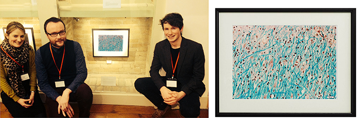
The image is a light microscopic image of part of the superior frontal gyrus, a region in the frontal lobe of the human brain, of an individual with Primary-Progressive Multiple Sclerosis (MS). Luxol fast blue (LFB) stain has been used to demonstrate the intricate pattern of myelin observed under light microscopy. Using this stain myelin fibres appear blue. LFB has been used in combination with a common hematoxylin and eosin stain. Hematoxylin is a purple dye that stains nuclear material, while eosin is an orange to red dye that stains the cytoplasm. Myelin is a substance rich in lipids and proteins that insulates nerve fibres, allowing efficient transmission of nerve impulses. In MS, an abnormal immune reaction is believed to initiate an attack on the myelin, resulting in its destruction and the appearance of lesions (bare spots and scarred areas along the nerve) throughout the brain. This images shows a region of the human brain that is lesion free, and hence we can observe the extensive network of myelin.
Grant support from the Health Research Board (HRB) is acknowledged. Tissue samples were supplied by the Multiple Sclerosis Society Tissue Bank, funded by the Multiple Sclerosis Society of Great Britain and Northern Ireland, registered charity 207495.
RTE Six One News covered the event see video link here
Department of Anatomy and Neuroscience
Anatamaíocht agus Néareolaíocht
Contact us
Room 2.33, 2nd Floor, Western Gateway Building, University College, Cork, Ireland