In This Section
- Home
- About the Department
- Welcome from Head of Department of Anatomy and Neuroscience
- A History of the Department
- A history of the Department; The early years to the 1980s
- A history of the Department; The move from the Windle Building to BSI and WGB
- UCC Professors of Anatomy and Heads of Department
- The development of the UCC HUB
- Current students, recent research graduates and awards
- Useful Links
- People and Phonebook
- Study Anatomy
- Study Neuroscience
- Research
- Neural circuitry underlying Neuropsychiatric and Neurological Disorders 2026
- Neurogastroenterology 2026
- Developmental Neuroscience and Regeneration 2026
- Neurodegeneration 2026
- Neuroinflammation 2026
- Neuroprotection and Therapeutics 2026
- Neuroproteomics and Molecular Psychiatry 2026
- Anatomy Education Research 2026
- Research Facilities 2026
- Postgraduate Research Programmes 2026
- UCC Anatomical Donations
- Biosciences Imaging Centre
- News & Events
- 2025 News Archive
- News Archive 2024
- News Archive 2023
- News Archive 2022
- News Archive 2021
- News Archive 2020
- News Archive 2019
- News Archive 2018
- News archive 2017
- News Archive 2016
- News Archive2015
- News Archive 2014
- News Archive 2013
- News Archive 2012
- News Archive 2011
- BRAIN AWARENESS WEEK 2023
- Department Events and Conferences
- Seminar series 2019_2020
- photo galleries
- Narrowing the void Conference 2023
- Photos of BSc Medical and Health Sciences Mentoring launch 2022
- International Women's Day 2023
- 2023 BRIGHT FUTURES - Celebrating our researchers
- 2023 UCC Futures - Future Ageing & Brain Sciences
- Recent Graduations July 2023
- Anatomy and Neuroscience Top 100 Anatomy Physiology 2023
- BRAIN AWARENESS WEEK 2023 FUN AND GAMES EVENT
- Medical and Health Sciences First year class 2023
- 2023 Brain Awareness week Scientific discussion photo gallery
- World Anatomy Day 2023
- BSc MHS MENTORING PROGRAMME 2023
- BSc Medical and Health Sciences Graduation 2023
- BSc Neuroscience Graduation Photo Gallery 2023
- Dr Kathy Quane Nov 2023
- THANKSGIVING PHOTOS 2012
- Photo Gallery: Society of Translational Medicine Careers Fair 2023
- Photo Gallery:2023 TRAIN AWARDS
- Photo Gallery:2024 Creative Week St Joseph's NS
- Photo Gallery: Department of Anatomy and Neuroscience Thanksgiving Service 2024
- Photo Gallery: Professor Aideen Sullivan farewell party
- Photo Gallery: Irish Pain Society Annual Scientific Meeting Cork 2023
- Photo Gallery: 2024 Medical and Health Sciences Graduation
- Photo Gallery: Medical and Health Sciences Meet and Greet 2024
- Photo Gallery: 2024 BSC NEUROSCIENCE Graduation
- Photo Gallery: 2025 INTERNATIONAL WOMEN'S DAY
- Photo Gallery: 2025 BSc Neuroscience class and staff
- Photo Gallery: 2025 BRAIN CONNECTIONS
- BSc Neuroscience Graduation Photo Gallery 2025
- World Anatomy Day 2025
- UCC Learning and Teaching Showcase 2025
- MSc Human Anatomy Graduation Photo Gallery 2025
- Department of Anatomy & Neuroscience DELTA AWARD 2026
- Thanksgiving Service 2026
- BSc Neuroscience Class photos 2026
- Flame Sculpture by Alexandra Wejchert
- FLAME LABORATORY GALLERY
- Narrowing the Void Conference 2023
- Department of Anatomy and Neuroscience Contact Us
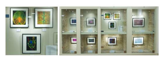
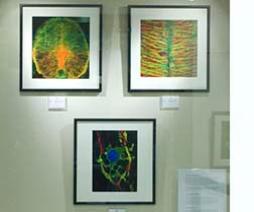
Anatomy & Neuroscience exhibit at the 'It’s a Beautiful World’ exhibition
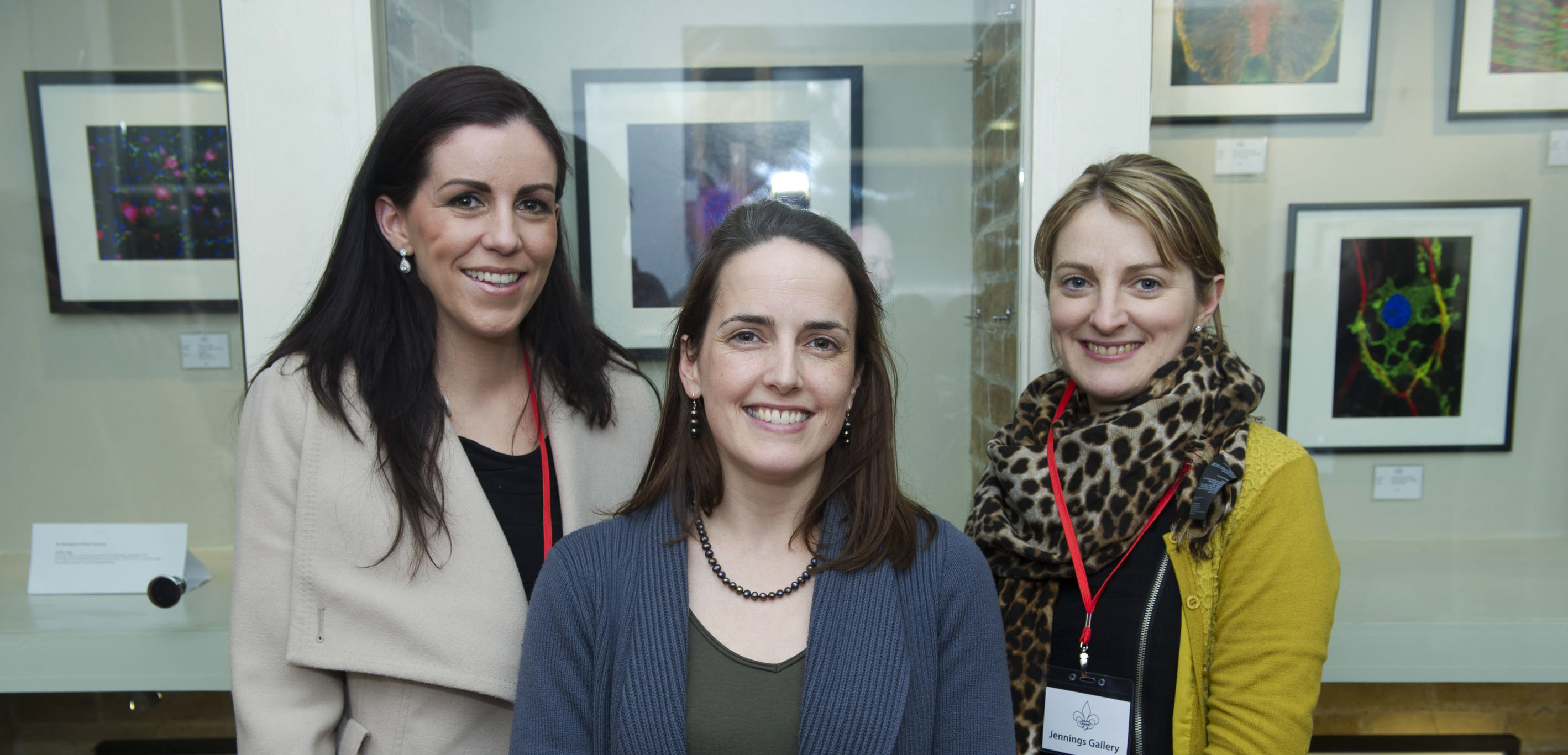
'It’s a Beautiful World’ exhibition opened on Thursday 20th February 2014 in the Jennings Gallery, College of Medicine and Health, Brookfield Health Sciences Complex, UCC.
The exhibition features a range of scientific images from the world of Medicine and Science and includes electron-microscopy images, anatomical drawings, baby brainwaves and neuroscience images. There is also a Natural Sciences section, featuring images from nature and life drawings. The exhibition includes photographs, paintings, drawings and other media. ‘Every day we see beautiful images in our work as scientists and healthcare workers. It’s a Beautiful World is a celebration of the beauty of Science and Nature.
The artists are all staff and students from the College of Medicine and Health at UCC (Dentistry, Medicine, Nursing and Midwifery, Pharmacy, Clinical Therapies) and UCC Research Groups and Teaching Hospitals.
The department of Anatomy and Neuroscience are well represented at the exhibition. Images from the students of Dr Yvonne Nolan, Dr André Toulouse, Dr Eric Downer and Dr Kieran McDermott are among the exhibits from the department of Anatomy and Neuroscience.
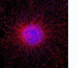 Becoming Neurons
Exhibited by Louise Collins, Suzanne Crotty and Yvonne Nolan, Department of Anatomy and Neuroscience
The birth of new neurons from neural stem cells occurs in the embryo and continues throughout adulthood in discreet regions of the mammalian brain. Neural stem cells group together in ball-like clusters called neurospheres and can be induced to give rise to neurons. Neurospheres are of great interest therapeutically as they could theoretically be used to generate and replace the neurons that are lost in traumatic brain injury and in neurodegenerative diseases such as Parkinson’s disease, Alzheimer’s disease and multiple sclerosis.
Image: This neurosphere was prepared from neural stem cells extracted from an embryonic rat midbrain. After allowing the cells to proliferate and expand for 7 days, they were then imaged with confocal microscopy. Cells differentiated to neurons are labeled red with an antibody to βIII-tubulin. All nuclei are stained blue with bisbenzamide.
We acknowledge Dr. Frank van Pelt for artistic input.
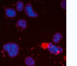 Au coeur de la cellule
Exhibited by André Toulouse, Julie Roussel and Guy Rouleau, Department of Anatomy and Neuroscience
Polyglutamine diseases are a group of neurological movement disorders including Huntington 's disease and several spinocerebellar ataxias. We created a cellular model to study abnormal protein production in
these diseases and while characterizing the cells under fluorescence microscopy, we observed this heart shaped protein inclusion at the periphery of a nucleus.
André Toulouse is a Lecturer and researcher in the Department of Anatomy and Neuroscience. His research gives him the opportunity to capture quirky moments around the life and death of cells. Dr Toulouse would
like to thank Julie Roussel and Guy Rouleau for their help.
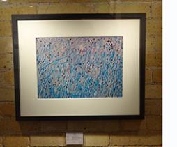
 Insulating nerve fibres, Making nerve cells, Spinal cord scaffolding
Exhibited by Kieran McDermott and Janelle Pakan, Department of Anatomy and Neuroscience
Insulating nerve fibres. This a very high magnification image of a brain cell taken with a con focal microscope which allows you to look deep into the brain tissue. The cell is called an oligodendrocyte and is the cell that makes the special insulating material (coloured green here) which surrounds nerve fibres (coloured red here ). The cell nucleus is coloured blue. The special staining procedure that was used here ensures that only these particular features are visible and the rest of the brain tissue is invisible. The cell is magnified over
a thousand times .
Making nerve cells. This is a very high magnification image of a tiny piece of the developing spinal cord taken with a confocal microscope which allows you to look deep into the spinal cord tissue. The sl it in the centre is the future central canal of the spinal cord and it is surrounded by a large number of spinal cord stem cells . These cells use the radially arranged filam ents (coloured green here) to migrate outward to increase the thickness of the cord during its development. The special staining procedure that was used here ensures that only these particular features are visible and the rest of the brain tissue is invisible. The cells are magnified over a thousand times.
Spinal cord scaffolding This is an image of a slice through the entire spinal cord during its embryonic development and is taken with a confocal microscope which allows you to look deep into the spinal cord tissue. The vertical slit in the centre is the future central canal of the spinal cord. The green coloured fibres form a very important radial scaffold which spans the thickness of the spinal cord and which spinal cord stem cells use to move on as the spinal co rd grows and expands in early life. Th e red coloured parts in the lower half of the spinal cord represent cells which have changed and developed into a more mature type of spinal cord cell.
Kieran McDermott is a Senior Lecturer in Anatomy and Neuroscience at UCC and Director of the BioSciences Imaging Centre. He has been interested for many years in the cellular and molecular mechanisms which allow the nervous system of mammals to develop, from a very primitive tube in the embryo, into the extremely complex adult organ. He has always employed advanced microscopy in his research and together with members of his laboratory, has received several imaging and microscopy awards. Dr McDermott's laboratory is interested in using advanced microscopy techniques to promote our understanding of the highly complex nature of brain and spinal cord development and in how such new knowledge can provide useful clues in the search for novel therapies for nervous system disease and injury. Dr McDermott believes that scientific images such as these serve to bridge the gap between science and art and can help to foster public interest in the scientific endeavour.
Janelle Pakan completed a Post-Doctoral Fellowship in the Department of Anatomy and Neuroscience at UCC with Dr Kieran McDermott. Her research looked at radial glial cell migration and differentiation in the developing spinal cord using two-photon imaging of living tissue. Janelle has recently taken up a Postdoctoral Fellowship at the University of Edinburgh.
 Myelin is getting on my nerves
Exhibited by Eric Downer, Department of Anatomy and Neuroscience
This is a light microscopic image of part of the frontal lobe of the human brain of an individual with Multiple Sclerosis (MS). Luxol fast blue stain has been used to demonstrate the intricate pattern of myelin fibres which are stained blue. A purple dye stains the nucleus of all cells. Myelin insulates nerve fibres , allowing efficient transmission of nerve impulses. In MS, myelin is lost, resulting in impaired nerve impulses and resultant clinical symptoms. This image was collated as part of a HRB-funded summer scholarship awarded to Mr. Richard Magee (currently a 4th year Neuroscienc e BSc student at UCC) to conduct research with Dr. Downer in collaboration with Dr. Yvonne Nolan (Dept. Anatomy and Neuroscience, UCC). Grant support from the Health Research Board (HRB) is acknowledged. Tissue samples were supplied by the Multiple Sclerosis Society Tissue Bank, funded by the Multiple Sclerosis Soc iety of Great Britain and Northern Ireland.
Dr. Eric Downer is a Lecturer in Anatomy and Neuroscience at UCC. His research interest is in Neuroimmunology and he leads a research group that targets the role of the innate immune system in Multiple Sclerosis.
RTE Six One News covered the event see video link here
Department of Anatomy and Neuroscience
Anatamaíocht agus Néareolaíocht
Contact us
Room 2.33, 2nd Floor, Western Gateway Building, University College, Cork, Ireland