- Home
- Staff Profiles & Phone Book
- About the Department
- Study Anatomy
- Study Neuroscience
- Research
- UCC Anatomical Donations
- Biosciences Imaging Centre
- BSc Medical and Health Sciences
- News & Events
- BRAIN AWARENESS WEEK 2023
- NEWS ARCHIVE 2023
- News Archive 2022
- News Archive 2021
- News Archive 2020
- News Archive 2019
- News Archive 2018
- News archive 2017
- Recent Publications
- News Archive 2016
- News Archive2015
- News Archive 2014
- News Archive 2013
- News Archive 2012
- News Archive 2011
- Department Events and Conferences
- Seminar series 2019_2020
- photo galleries
- Department of Anatomy and Neuroscience Contact Us
Image of the month image details
February 2017
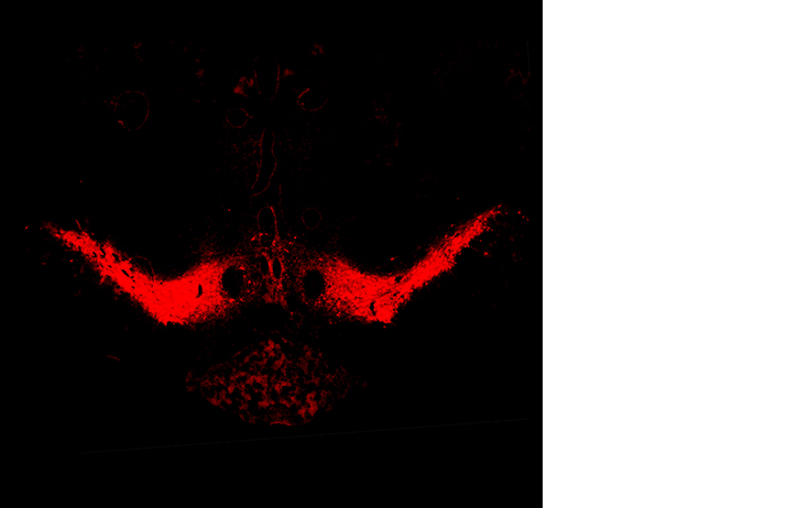
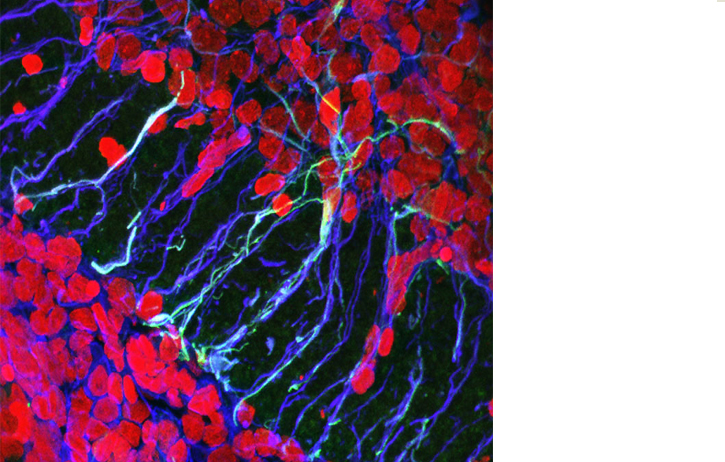
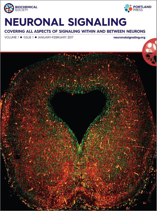 February 2017: "The heart of the brain”: the developing midbrain
Submitted by: Dr Shane V Hegarty, Dr. Gerard O'Keeffe and Professor Aideen Sullivan, Department of Anatomy and Neuroscience, UCC, Ireland.
Image shows a coronal section of embryonic day 16.5 mouse midbrain, immunohistochemically stained for tyrosine hydroxylase and DAPI. Tyrosine hydroxylase is used to identify midbrain dopaminergic neurons, while DAPI shows the nuclei of cells in this brain region. Dr. Shane Hegarty, Dr. Gerard O'Keeffe and Professor Aideen Sullivan research the molecular signals regulating the development of midbrain dopaminergic neurons with the aim of translating this information into novel therapies for Parkinson's disease, a common neurodegenerative disorder in which this neuronal population is progressively degenerated.
This image was selected for the cover of the first issue of Neuronal Signaling a bi monthly journal publishing high-quality molecular and cellular neuroscience research. Neuronal Signaling spans a variety of neuroscientific disciplines, from signaling pathways involved in nervous system development through to neurodegeneration, synaptopathies, psychiatric disorders and other pathologies.
In this, the first issue of Neuronal Signaling, Aideen Sullivan, the Editor-in-Chief, presents the journal's aims and scope in her introductory Editorial.
January 2017
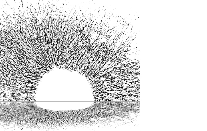
January 2017: A Winter Sunset
Submitted by: Dr Gerard O'Keeffe
Dr O'Keeffe is following on his theme of finding art under the Microscope. For December Dr O'Keeffe has edited an image of a explant culture of a sympathetic ganglion. By thresholding the image and reflecting the structure he has created a seasonally atmospheric Sunrise Sunset image.
November 2016
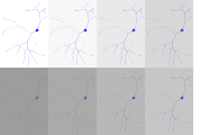
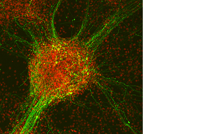
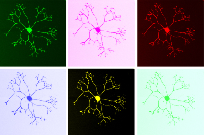 November 2016: Fifty shades of Grey matter
Submitted by: Dr Gerard O'Keeffe
Parkinson’s is a neurodegenerative disorder characterised by the degeneration of nerve cell projections (axons) and later the death of nerve cells (neurons) from the central and peripheral nervous system. We are studying the molecular mechanisms that promote neuronal survival and growth to identify new drugs and drug targets for Parkinsons. Shown here is one single sympathetic neuron which grow and branch extensively in culture which we use to study affected axons. The title of this image represents the diversity in symptoms in Parkinson’s and the many types of neurons affected by the disorder.
October 2016
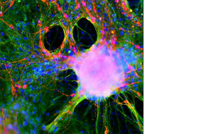
October 2016: A Scream for Halloween
Submitted by: Dr Gerard O'Keeffe
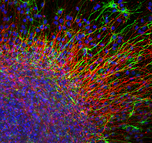
An image of a culture of neurons derived from neural precursor cells - dopamine neurons are shown in red, all neurons are shown in green and nuclei are shown in blue, seems like any other image taken in a day in the lab. But as Halloween approaches strange images begin to appear and Dr Gerard O'Keefe is unsure if this is the revisiting of Edvard Munch's most famous work, “The Scream” or is it something more omonious.... perhaps it is in fact Ghostface from the Scream films. Happy Halloween!!
September 2016
September 2016: More than Neurons feeling the blues
Submitted by: Dr Gerard O'Keeffe
According to the World Health Organisation, depression is a common mental disorder. Globally, an estimated 350 million people of all ages suffer from depression. It is the leading cause of disability worldwide, and is a major contributor to the overall global burden of disease. The image represents the many different emotions and their severity reflected in grey, overlaid on a neuron to represent the need to better understand the neurological and psychological origins of this disorder.
August 2016
"cellular factories". Each image shows a single CHO cell that has been engineered to overexpress a neurotrophic factor for cell transplantations approaches in Parkinson's disease.
July 2016
"An abstract scientific abstract" - its a mixed culture of neurons from the embryonic rat brain showing dopamine neurons in red, neurons in green and cell nuclei in blue.
June 2016
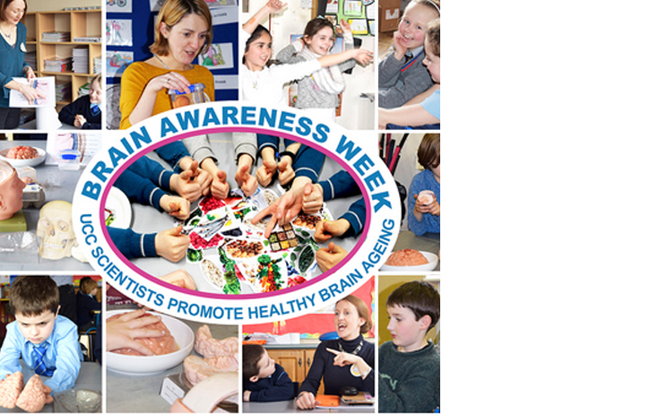
“Tús Maith Leath na hOibre” During Brain Awareness Week, March 14-20, 2016 a group of UCC neuroscientists engaged children from five local primary schools Glounthaune, Greenmount, Glasheen Girls, Glasheen Boys and St Finbarres in fun activities to promote the importance of brain health during childhood.
May 2016
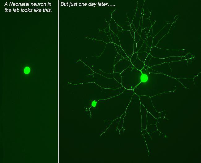 May 2016: Aren't the Neurons growing up so quickly
Submitted by: Dr Gerard O'Keeffe
Images of neuron cultures from the neonatal nervous system - beautiful and elegant growth of axonal projections captured under the microscope. The remarkable complexity and speed of growth at this stage of development is impressive.
April 2016

April 2016: April is Parkinson's Awareness Month!
Submitted by: Professor Aideen Sullivan
The dance by people with Parkinson’s and researchers was recorded as a digital video by Brid Corcoran, with the movements and interaction within this digital art piece exemplifying the spirit of the The ‘Parkinson’s Community’ meeting which was organised by Dr Shane Hegarty and Professor Aideen Sullivan of the Department of Anatomy and Neuroscience, UCC, as part of the BRAINTALK project (www.ucc.ie/en/braintalk).
The purpose of the meeting was to bring together People with Parkinson's, Parkinson's researchers and clinicians, to create an interconnected Parkinson's Community in Ireland. Over 200 participants, the majority of whom were People with Parkinson’s and their carers, gathered in the Glucksman Gallery for a variety of presentations by Parkinson’s advocacy groups, People with Parkinson’s, therapists, neurologists and neuroscientists.
To conclude the event, several People with Parkinson’s paired up with neuroscientists for a set-dancing session directed by Pat O’Dea. This dance was as a dialogue between those affected by Parkinson’s and those working to develop new therapies, and thus a fitting finale to the day.
Link to website for further details.
http://www.ucc.ie/en/anatomy/news/fullstory-598438-en.html
http://www.ucc.ie/en/braintalk/
April is Parkinson's Awareness Month! link
http://www.pdf.org/en/spring10_april_parkinson_awareness_month
March 2016
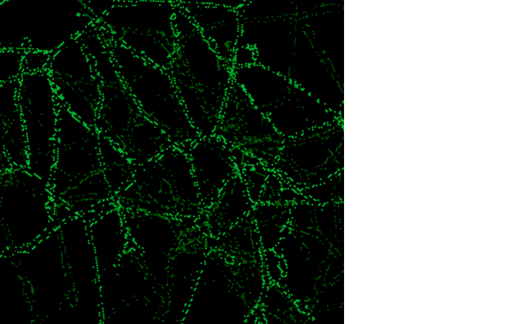
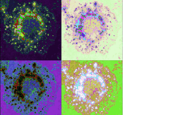
Title: ‘The ghost in the machine.’
Description: Axonal networks are crucial for brain health and disruption of these networks are seen in many brain diseases and disorders. The Image shows a layered image made with beta-III stained axons from individual neurons layered on top of one another. The colour was adjusted in each layer to represent the three dimensional nature of axonal networks in vivo.
February 2016
February 2016: "Expression of NADPH diaphorase (a marker of nitric oxide synthase (NOS) enzyme activity) in rat cortical neurons"
Submitted by: Submitted by Dr Eoin Sherwin
Dr Eoin Sherwin is a post-doctoral researcher with the APC Microbiome institute. His research focuses on the development of live biotherapeutics for the treatment of autistic spectrum disorders.
The image depicts rat cortical neurons that has been stained with NADPH diaphorase. NADPH diaporase staining is indicative of cells that are actively producing the free radical gas, nitric oxide, via the enzymatic actions of nitric oxide synthase."
January 2016
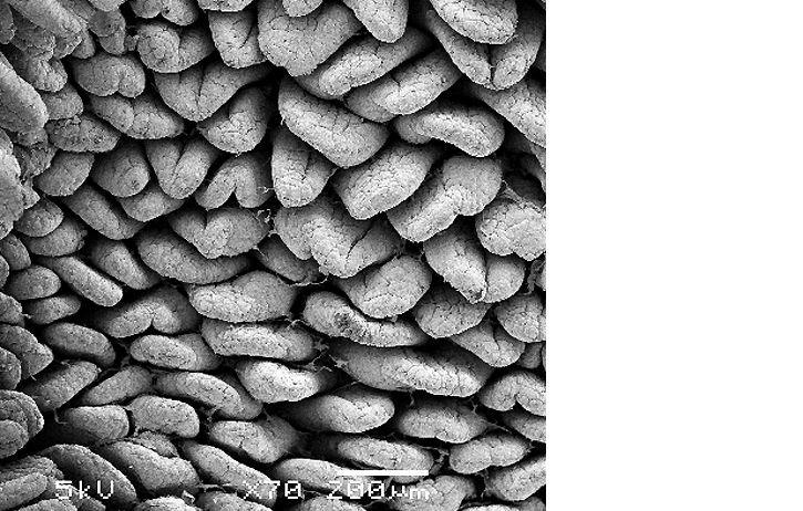
January 2016: Rat intestine
Submitted by: Suzanne Crotty
Image shows the internal surface of a rat intestine which has been prepared by sputter coating the surface with gold and imaged using a Jeol JSM 5510 Scanning Electron Microscope.
Suzanne Crotty is a Senior Technical Officer in the Department of Anatomy and Neuroscience, UCC, and looks after the day to day running of the BioSciences Imaging Centre.
Suzanne graduated with a B.Sc. (Hons) and M.Sc. in Neuroscience from University College Cork. She has expertise in electron, light and fluorescence microscopy including wide-field, confocal and two-photon microscopy. Suzanne has a strong research background in the preparation of biological specimens for microscopy, including scanning and transmission electron microscopy.
The Bioscience Imaging Centre provides a microscopy service for staff and researchers throughout the university, and also investigators from other universities and from industry. Services provided at the BioSciences Imaging Centre include a wide range of microscopy techniques, specimen preparation (of biological or materials samples) and microscopy training.
December 2015
December 2015: ‘Cluster Birth’
Submitted by: Professor Aideen Sullivan
This image was recently exhibited as part of the BRAINTALK Parkinson’s Community Meeting and Art Exhibition, held in the Glucksman Gallery University College Cork. This event was organised by Dr Shane Hegarty and Professor Aideen Sullivan of the Department of Anatomy and Neuroscience, UCC, as part of the BRAINTALK project (www.ucc.ie/en/braintalk).
The purpose of the meeting was to bring together People with Parkinson's, Parkinson's researchers and clinicians, to create an interconnected Parkinson's Community in Ireland. Over 200 participants, the majority of whom were People with Parkinson’s and their carers, gathered in the Glucksman Gallery for a variety of presentations by Parkinson’s advocacy groups, People with Parkinson’s, therapists, neurologists and neuroscientists.
‘Parkinson’s Community’ art exhibition was launched after the meeting. This included paintings made by People with Parkinson’s in an ‘Exploring Parkinson’s with Art’ workshop , as well as photomicrographs showing the ongoing scientific work by Parkinson’s researchers in the Department of Anatomy and Neuroscience, UCC.
For full details see BRAINTALK website http://www.ucc.ie/en/braintalk/
November 2015
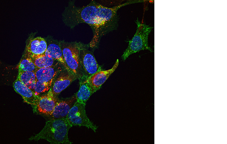
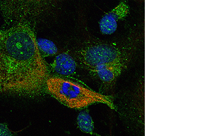
November 2015: Co-expression of the central “hunger” receptor, ghrelin, also called growth hormone secretagogue receptor (green) and the serotonin 2C (5HT2C) receptor (red) in a culture of human embryonic kidney cells.
Submitted by: Dr Harriet Schellekens
Dr. Harriët Schellekens is a Lecturer in the department of Anatomy & Neuroscience and a Principal Investigator with APC Microbiome Institute and Food for Health Ireland. Her research focusses on the neuronal circuitry underlying the complex relationship between stress, mood and food intake. In particular, her work involves the pharmacological targeting of G-protein coupled receptors (GPCRs) expressed in the brain. Recently, she has identified a novel heterodimer between two key GPCRs important in the central regulation of feeding behaviour, namely, the ghrelin or GHS-R1a receptor and the 5-HT2C receptor (Schellekens et al., JBC, 2013; Schellekens et al., ACS Chemical Neuroscience, 2015). These findings have uncovered a novel mechanism of serotonin-mediated attenuation of the ghrelin receptor, which is poised to have significant impact on the future pharmacological targeting of the ghrelin receptor in the homeostatic regulation of body weight as well as hedonic appetite signalling, which both play a significant role in the development of obesity.
The image depicts human embryonic kidney (Hek) cells with stable expression of the ghrelin receptor (green). Cells also express the 5HT2C receptor (red) after lentiviral transduction. Co-localized receptor expression and possible heterodimerization is indicated by yellow colour overlap. Cell nuclei were stained with bisbenzamide (blue). The image was taken using confocal microscopy.
Further reading:
Schellekens, H., De Francesco, P.N., Kandil, D., Theeuwes, W.F., McCarthy, T., van Oeffelen, W.E.P.A., Perelló, M., Giblin, L., Dinan, T.G., Cryan, J.C., Ghrelin’s orexigenic effect is modulated via 1 a serotonin 2C receptor interaction, ACS Chemical Neuroscience, 2015.
Schellekens, H., van Oeffelen, W.E.P.A., Dinan, T.G., Cryan, J.F., Promiscuous Dimerization of the Growth Hormone Secretagogue Receptor (GHS-R1a) Attenuates Ghrelin-mediated Signaling. J Biol Chem, 2013. Jan; 288(1): 181-91.
October 2015
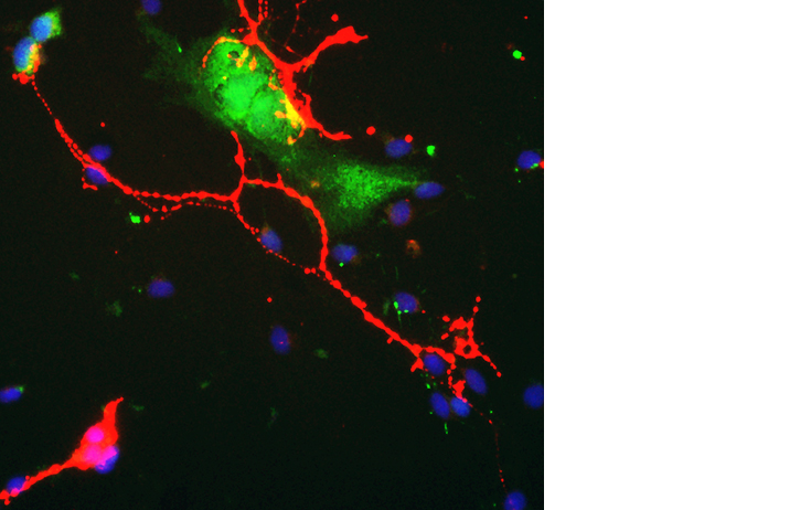
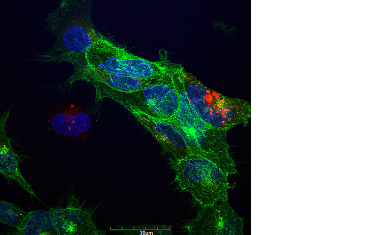
October 2015: Embryonic ventral midbrain dopaminergic neuron (red) synpases on a transfected non-dopaminergic neuron (green) expressing green fluorescent protein.
Submitted by: Shane V. Hegarty Post-Doctoral Research Fellow, Department of Anatomy and Neuroscience.
Dr Shane Hegarty is a BSc and PhD graduate from the Department of Anatomy and Neuroscience, UCC, who recently began an Irish Research Council Post-Doctoral Research Fellowship under the supervision of Dr Aideen Sullivan and Dr Gerard O’Keeffe. Their research investigates the role of BMP-Smad signalling in ventral midbrain dopaminergic neuron development.
Transfection allows the introduction of specific sequences of nucleic acids, e.g. DNA or RNA, into a desired cell type, and is generally employed to alter the expression levels of a protein of interest. Transfection of primary neurons enables the investigation of the functional roles of various target genes in neuronal development and survival. These transfected neurons can be visualised through the ectopic expression of fluorescent proteins, such as green fluorescent protein (GFP).
The image depicts an embryonic ventral midbrain dopaminergic neuron (red) which has formed synapses on a transfected non-dopaminergic neuron (green), expressing green fluorescent protein, in a culture of the embryonic ventral midbrain following four days of differentiation. Synapses are the point of electrochemical communication between neurons. Embryonic day 14 ventral midbrain cells were transfected via electroporation with specific plasmid(s), and subsequently differentiated using serum proteins. A transfected glial cell (green) also appears to be in contact with the processes of another dopaminergic neuron (red). Cellular nuclei were stained with bisbenzamide (blue). The image was taken using an inverted fluorescence microscope at 200x magnification.
This image was recently exhibited as part of the BRAINTALK Parkinson’s Community Meeting and Art Exhibition, held in the Glucksman Gallery University College Cork. This event was organised by Dr Shane Hegarty and Professor Aideen Sullivan of the Department of Anatomy and Neuroscience, UCC, as part of the BRAINTALK project (www.ucc.ie/en/braintalk).
The purpose of the meeting was to bring together People with Parkinson's, Parkinson's researchers and clinicians, to create an interconnected Parkinson's Community in Ireland. Over 200 participants, the majority of whom were People with Parkinson’s and their carers, gathered in the Glucksman Gallery for a variety of presentations by Parkinson’s advocacy groups, People with Parkinson’s, therapists, neurologists and neuroscientists.
‘Parkinson’s Community’ art exhibition was launched after the meeting. This included paintings made by People with Parkinson’s in an ‘Exploring Parkinson’s with Art’ workshop , as well as photomicrographs showing the ongoing scientific work by Parkinson’s researchers in the Department of Anatomy and Neuroscience, UCC.
For full details see BRAINTALK website http://www.ucc.ie/en/braintalk/.
September 2015
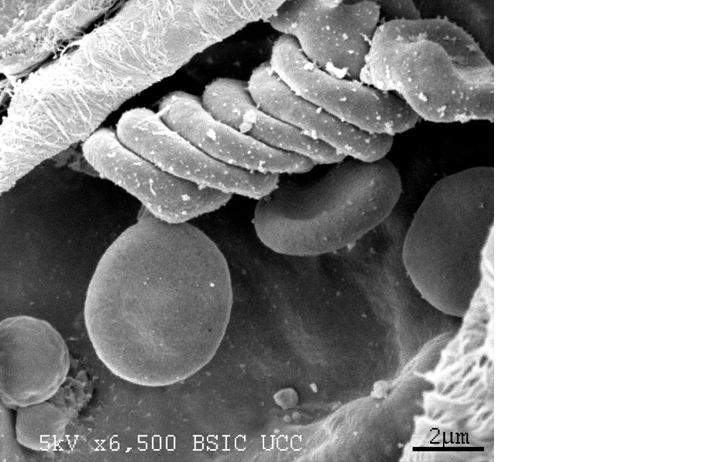
September 2015: Red blood cells on rat muscle sample
Submitted by: Suzanne Crotty
Image shows the surface of an exposed rat muscle which has been prepared by sputter coating the surface with gold and imaged using a Jeol JSM 5510 Scanning Electron Microscope.
Suzanne Crotty is a Senior Technical Officer in the Department of Anatomy and Neuroscience, UCC, and looks after the day to day running of the BioSciences Imaging Centre.
Suzanne graduated with a B.Sc. (Hons) and M.Sc. in Neuroscience from University College Cork. She has expertise in electron, light and fluorescence microscopy including wide-field, confocal and two-photon microscopy. Suzanne has a strong research background in the preparation of biological specimens for microscopy, including scanning and transmission electron microscopy.
The Bioscience Imaging Centre provides a microscopy service for staff and researchers throughout the university, and also investigators from other universities and from industry. Services provided at the BioSciences Imaging Centre include a wide range of microscopy techniques, specimen preparation (of biological or materials samples) and microscopy training.
August 2015
August 2015: ’Wings in Motion’
Submitted by: Erin Dolan, Yvonne Nolan, Aideen Sullivan
Erin Dolan is a PhD student in the Department of Anatomy and Neuroscience under the supervision of Prof Aideen Sullivan and Dr Yvonne Nolan, and is funded by the Molecular Medicine Ireland Clinical and Translational Research Scholarship.
Parkinson’s disease is the second most common neurodegenerative disorder in the world, and is characterised by the death of dopaminergic neurons in the nigrostriatal pathway of the brain, leading to a significant decrease in the neurotransmitter dopamine. The resulting loss of dopaminergic transmission in this brain region leads to the primary motor symptoms associated with the disease, including a fine tremor at rest, bradykinesia, postural instability and rigidity. The current mainstay of treatment is simply to replace the lost dopamine, however therapy is difficult to optimise and the effects wear off over time.
This image, which depicts an intact rodent substantia nigra with the characteristic “wings” of the pars reticulata, is stained with tyrosine hydroxylase, a marker specific to dopaminergic neurons. Novel therapeutic strategies in Parkinson’s disease aim to either replace the lost dopaminergic neurons (neurorestoration) or to protect the surviving neurons from further insult (neuroprotection), and evaluating the integrity of the nigrostriatal system through tyrosine hydroxylase staining is one of the most effective methods used in current research. This image was taken at 4x magnification on an Olympus BX53 upright microscope and processed using Cellsens software
This image was recently exhibited as part of the BRAINTALK Parkinson’s Community Meeting and Art Exhibition, held in the Glucksman Gallery University College Cork. This event was organised by Dr Shane Hegarty and Professor Aideen Sullivan of the Department of Anatomy and Neuroscience, UCC, as part of the BRAINTALK project (www.ucc.ie/en/braintalk).
The purpose of the meeting was to bring together People with Parkinson's, Parkinson's researchers and clinicians, to create an interconnected Parkinson's Community in Ireland. Over 200 participants, the majority of whom were People with Parkinson’s and their carers, gathered in the Glucksman Gallery for a variety of presentations by Parkinson’s advocacy groups, People with Parkinson’s, therapists, neurologists and neuroscientists.
‘Parkinson’s Community’ art exhibition was launched after the meeting. This included paintings made by People with Parkinson’s in an ‘Exploring Parkinson’s with Art’ workshop , as well as photomicrographs showing the ongoing scientific work by Parkinson’s researchers in the Department of Anatomy and Neuroscience, UCC.
For full details see BRAINTALK website http://www.ucc.ie/en/braintalk/
July 2015
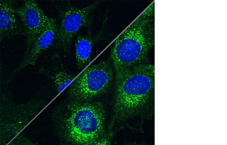
July 2015: ‘Switching on the Light’
Loss of the PINK1 protein function causes rare inherited forms of Parkinson's disease. By working out how PINK1 functions, researchers can learn how PINK1 protects the brain from Parkinson’s “switching the light” on better ways to treat, diagnose and hopefully in the long term prevent Parkinson’s disease.
Submitted by: Rachel Furlong, Cora O’Neill
Rachel Furlong is a BSc graduate from the Department of Anatomy and Neuroscience, UCC. She recently obtained an MSc in Molecular Cell Biology with Bioinnovation from the School of Biochemistry and Cell Biology UCC, conducting a research project in Parkinson’s disease supervised by Dr. Cora O’Neill. Rachel was awarded an Irish Research Council PhD postgraduate scholarship in 2015, and began her PhD under the supervision of Dr Cora O'Neill and Prof Aideen Sullivan in October 2015. Her PhD research project is investigating the mechanisms by which PINK1 protects against Parkinson’s disease: to develop better biomarkers and treatment targets for the disease.
The image above shows cells with no PINK1 (left of image) have reduced levels of a key signalling protein (labelled in green) in the PI3-kinase/Akt pathway, a major signalling hub for cell survival and cellular health. In contrast healthy cells containing the PINK1 protein (right of image) have ample amounts of this key survival signalling protein. Our research indicates that the interaction of PINK1 protein and this key survival signalling pathway is important in balancing cell survival and cell death decisions in Parkinson's disease. By elucidating how PINK1 and this survival pathway interact we hope to apply this knowledge to better understand, diagnose and treat Parkinson’s."
This image was recently exhibited as part of the BRAINTALK Parkinson’s Community Meeting and Art Exhibition, held in the Glucksman Gallery University College Cork. This event was organised by Dr Shane Hegarty and Professor Aideen Sullivan of the Department of Anatomy and Neuroscience, UCC, as part of the BRAINTALK project (www.ucc.ie/en/braintalk).
The purpose of the meeting was to bring together People with Parkinson's, Parkinson's researchers and clinicians, to create an interconnected Parkinson's Community in Ireland. Over 200 participants, the majority of whom were People with Parkinson’s and their carers, gathered in the Glucksman Gallery for a variety of presentations by Parkinson’s advocacy groups, People with Parkinson’s, therapists, neurologists and neuroscientists.
‘Parkinson’s Community’ art exhibition was launched after the meeting. This included paintings made by People with Parkinson’s in an ‘Exploring Parkinson’s with Art’ workshop , as well as photomicrographs showing the ongoing scientific work by Parkinson’s researchers in the Department of Anatomy and Neuroscience, UCC.
For full details see BRAINTALK website http://www.ucc.ie/en/braintalk/.
June 2015
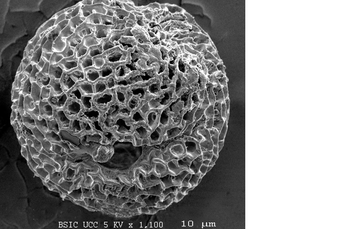
June 2015: Pollen grain
Submitted by: Suzanne Crotty
Image shows the surface of an pollen grain which has been prepared by sputter coating the surface with gold and imaged using a Jeol JSM 5510 Scanning Electron Microscope.
Suzanne Crotty is a Senior Technical Officer in the Department of Anatomy and Neuroscience, UCC, and looks after the day to day running of the BioSciences Imaging Centre.
Suzanne graduated with a B.Sc. (Hons) and M.Sc. in Neuroscience from University College Cork. She has expertise in electron, light and fluorescence microscopy including wide-field, confocal and two-photon microscopy. Suzanne has a strong research background in the preparation of biological specimens for microscopy, including scanning and transmission electron microscopy.
The Bioscience Imaging Centre provides a microscopy service for staff and researchers throughout the university, and also investigators from other universities and from industry. Services provided at the BioSciences Imaging Centre include a wide range of microscopy techniques, specimen preparation (of biological or materials samples) and microscopy training.
May 2015
May 2015:
Submitted by: Dr Harriet Schellekens
Dr. Harriët Schellekens is a Lecturer in the department of Anatomy & Neuroscience and a Principal Investigator with APC Microbiome Institute and Food for Health Ireland. Her research focusses on the neuronal circuitry underlying the complex relationship between stress, mood and food intake. In particular, her work involves the pharmacological targeting of G-protein coupled receptors (GPCRs) expressed in the brain. Recently, she has identified a novel heterodimer between two key GPCRs important in the central regulation of feeding behaviour, namely, the ghrelin or GHS-R1a receptor and the 5-HT2C receptor (Schellekens et al., JBC, 2013; Schellekens et al., ACS Chemical Neuroscience, 2015). These findings have uncovered a novel mechanism of serotonin-mediated attenuation of the ghrelin receptor, which is poised to have significant impact on the future pharmacological targeting of the ghrelin receptor in the homeostatic regulation of body weight as well as hedonic appetite signalling, which both play a significant role in the development of obesity.
The image depicts human embryonic kidney (Hek) cells with stable expression of the ghrelin receptor (green). Cells also express the 5HT2C receptor (red) after lentiviral transduction. Co-localized receptor expression and possible heterodimerization is indicated by yellow colour overlap. Cell nuclei were stained with bisbenzamide (blue). The image was taken using confocal microscopy.
April 2015
April 2015: Cluster of radial glial-like neural progenitors (red) generating immature neurons (green) in a culture of the embryonic rat ventral midbrain.
Submitted by: Shane V. Hegarty Post-Doctoral Research Fellow, Department of Anatomy and Neuroscience.
Dr Shane Hegarty is a BSc and PhD graduate from the Department of Anatomy and Neuroscience, UCC. He recently began an Irish Research Council Post-Doctoral Research Fellowship under the supervision of Dr Aideen Sullivan and Dr Gerard O’Keeffe in the Department of Anatomy and Neuroscience. Their research investigates the role of BMP-Smad signalling in ventral midbrain dopaminergic neuron development.
The differentiation of specific neuronal cell types from neural stem cells is a research area of major therapeutic interest, given the promising potential of such progeny to be utilised for cell replacement therapy, disease modelling and drug screening. The generation of ventral midbrain dopaminergic neurons from stem cells is of particular interest to the Parkinson’s disease field, due to the progressive loss of this neuronal population in this neurodegenerative disease.
The image depicts a cluster of vimentin-immunostained neural precursors (red), which have a radial glial-like morphology, in a culture of the embryonic ventral midbrain following one week of proliferation and two weeks of differentiation. Embryonic day 14 ventral midbrain neural stem cells were proliferated using mitogens, and subsequently differentiated using serum proteins. These neural precursors generate immature βIII-tubulin-immunostained neurons (green) during the second week of differentiation in vitro. Cellular nuclei were stained with bisbenzamide (blue). The image was taken using an inverted fluorescence microscope at 200x magnification.
This image was recently exhibited as part of the BRAINTALK Parkinson’s Community Meeting and Art Exhibition, held in the Glucksman Gallery University College Cork. This event was organised by Dr Shane Hegarty and Professor Aideen Sullivan of the Department of Anatomy and Neuroscience, UCC, as part of the BRAINTALK project (www.ucc.ie/en/braintalk).
The purpose of the meeting was to bring together People with Parkinson's, Parkinson's researchers and clinicians, to create an interconnected Parkinson's Community in Ireland. Over 200 participants, the majority of whom were People with Parkinson’s and their carers, gathered in the Glucksman Gallery for a variety of presentations by Parkinson’s advocacy groups, People with Parkinson’s, therapists, neurologists and neuroscientists.
‘Parkinson’s Community’ art exhibition was launched after the meeting. This included paintings made by People with Parkinson’s in an ‘Exploring Parkinson’s with Art’ workshop , as well as photomicrographs showing the ongoing scientific work by Parkinson’s researchers in the Department of Anatomy and Neuroscience, UCC.
For full details see BRAINTALK website http://www.ucc.ie/en/braintalk/
March 2015
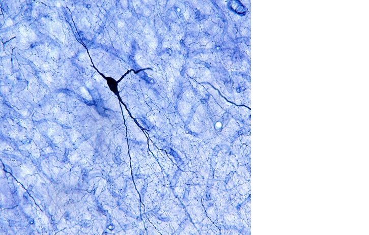
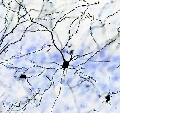 March 2015: Golgi-Cox stained pyramidal neuron located in the prefrontal cortex of a germ-free mouse
Submitted by: Pauline Luczynski M.Sc., Research Assistant , Neurogastroentorology Lab, Alimentary Pharmabiotic Centre.
Pauline Luczynski is a research assistant working with Prof. John Cryan's Neurogasterenterology research group, based in the Alimentary Pharmabiotic Centre. Her work studying neural remodelling along the gut-brain-microbiota axis was carried out in conjunction with the Department of Anatomy and Neuroscience.
The image depicts a Golgi-Cox stained pyramidal neuron located in the prefrontal cortex of a germ-free mouse. The Golgi-Cox stain randomly impregnates neurons, allowing the morphology of dendrites and spines to be studied using brightfield microscopy. The brain tissue was also stained with thionin (blue) to visualize cell layers. Brightfield image stacks were taken at 40x magnification using an OLYMPUS AX70 PROVIS microscope. Z-stacks were then merged using focus stacking software.
February 2015
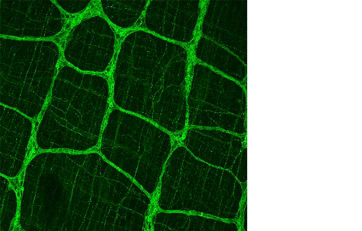 February 2015: Confocal Laser Scanning Microscope Image of myenteric plexus of the rat distal colon with a fine structure of nerve fibers.
Submitted by: Dr Anna V Golubeva, Post-Doctoral Researcher Neurogastroentorology Lab, Alimentary Pharmabiotic Centre.
Dr Anna Golubeva is a postdoctoral researcher in Professor John Cryan’s research group. based in the Alimentary Pharmabiotic Centre, her recent study on the intestinal innervation in prenatally stressed animals was carried out in conjunction with the Department of Anatomy and Neuroscience.
The attached image depicts the myenteric plexus of the rat distal colon with a fine structure of nerve fibers innervating underlying circular muscle layer. Colonic tissue was stained against choline acetyltransferase; the image was taken using confocal fluorescence microscopy.
January 2015
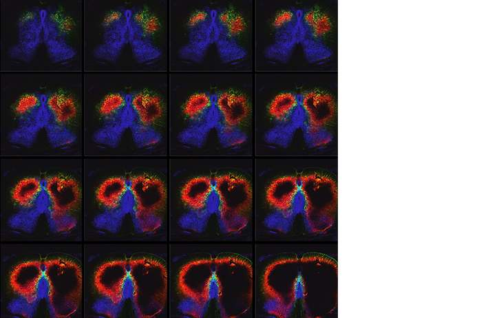
 January 2015: Confocal Laser Scanning Microscope Image projection of serial Z series sections.
Submitted by Dr Holly Greene, recent graduate from Department of Anatomy and Neuroscience.
TLX (orphan nuclear receptor tailless homologue) maintains adult neural stem cells in an undifferentiated, proliferative state. TLX (red) can silence glia-specific expression of the astrocyte marker GFAP (green) in neural stem cells, suggesting that transcriptional repression may be crucial in maintaining the undifferentiated state of these cells. Nuclei are stained blue with DAPI. Notice cell preparing to divide.
Click here for movie of Z series through the specimen
December 2014
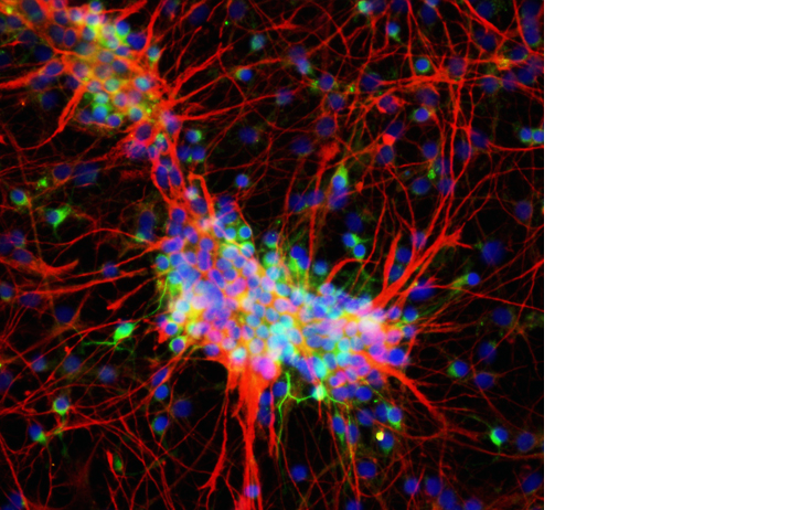
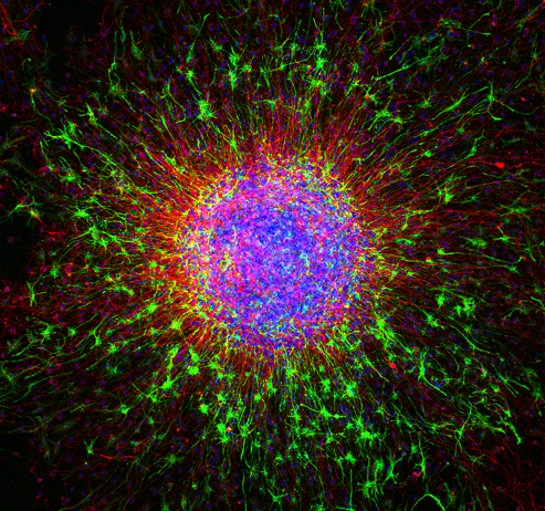
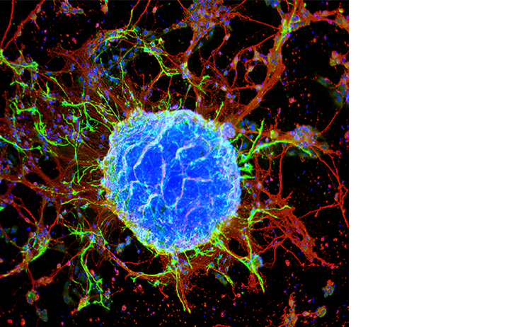
December 2014: Neuron Pipeline
Submitted by Yvonne Nolan, Louise Collins, Suzanne Crotty, Department of Anatomy and Neuroscience
When cultured with growth factors in vitro, neural stem cells can generate neurons (red), as well as the cells that support them— astrocytes (green) and oligodendrocytes. In culture, neural stem cells group together in ball-like clusters, called neurospheres (bottom left). Neurospheres are of great therapeutic interest because they have the potential to regenerate and replace neurons lost in traumatic brain injury and neurodegenerative diseases, such as Parkinson's disease, Alzheimer's disease, and multiple sclerosis.
Image: This neurosphere was prepared from neural stem cells extracted from an embryonic rat midbrain. After allowing the cells to proliferate and expand for 7 days, they were imaged with confocal microscopy. Cells differentiated to neurons are labeled red with an antibody to βIII-tubulin, whereas astrocytes are labeled green with an antibody to glial fibrillary acidic protein (GFAP), an intermediate filament specific to astrocytes. All nuclei are stained blue with bisbenzamide.
The image can be viewed at
http://www.cell.com/Cell_Picture_Show
November 2014
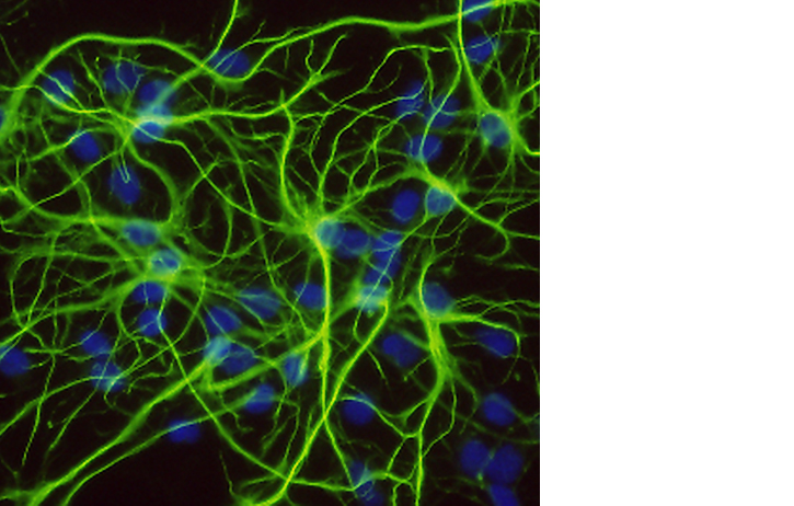
November 2014: Fluorescently labelled astrocytes in a mixed culture. Stained with GFAP add DAPI.
Submitted by Dr Danny Shanley, recent graduate Department of Anatomy and Neuroscience.
October 2014
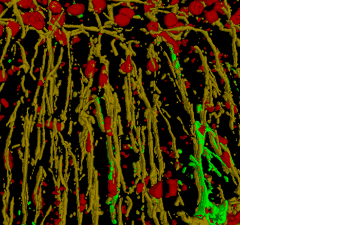
October 2014: 3D reconstruction of confocal image using Volocity Software
September 2014
September 2014: Confocal Laser Scanning Microscope montage of serial sections through a rat brain.
Submitted by Dr Conor O'Leary, recent graduate Department of Anatomy and Neuroscience.
August 2014
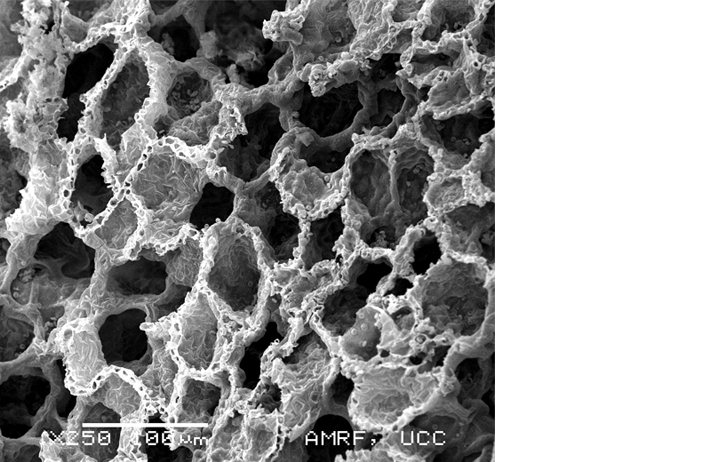
August 2014: Scanning Electron Micrograph of rat lung. Scale 100μm
Submitted by Ms Suzanne Crotty. Biosciences Imaging Centre.
Image shows the surface of an exposed rat lung which has been prepared by sputter coating the surface with gold and imaged using a Jeol JSM 5510 Scanning Electron Microscope.
Suzanne Crotty is a Senior Technical Officer in the Department of Anatomy and Neuroscience, UCC, and looks after the day to day running of the BioSciences Imaging Centre.
Suzanne graduated with a B.Sc. (Hons) and M.Sc. in Neuroscience from University College Cork. She has expertise in electron, light and fluorescence microscopy including wide-field, confocal and two-photon microscopy. Suzanne has a strong research background in the preparation of biological specimens for microscopy, including scanning and transmission electron microscopy.
The Bioscience Imaging Centre provides a microscopy service for staff and researchers throughout the university, and also investigators from other universities and from industry. Services provided at the BioSciences Imaging Centre include a wide range of microscopy techniques, specimen preparation (of biological or materials samples) and microscopy training.
July 2014
,SOX2(blue),July2014.jpg)
July 2014: Direct observation of cell division in BLBP-GFP transfected cells
Submitted by Dr Janelle Pakan and Dr Kieran Mc Dermott
Direct observation of cell division in BLBP-GFP transfected cells Confocal image of a single late telophase GFP+(green), SOX2+(blue), Ki67+(red) cell and several Ki67+SOX2+ cells, indicating preserved proliferative capacity and slice viability.
June 2014
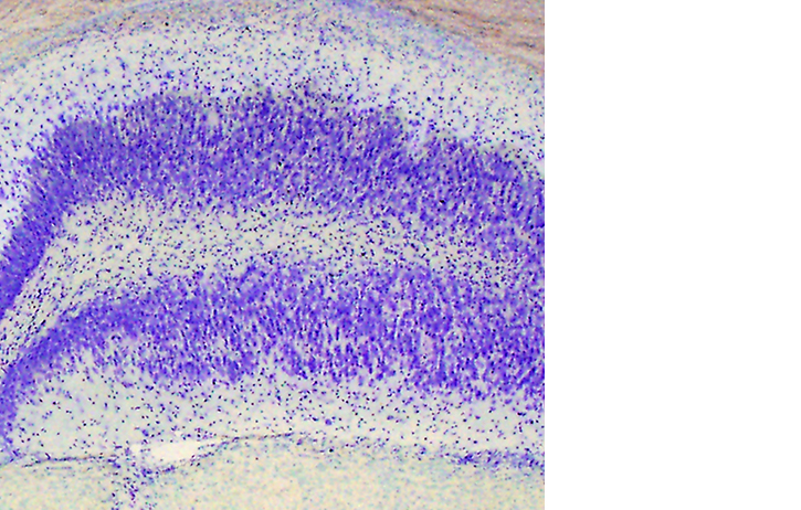
June 2014: Cresyl violet (nissl stain) image of the hippocampus from a mouse model of Temporal Lobe Epilepsy
Submitted by Elaine O’Loughlan Department of Anatomy & Neuroscience
Image shows severe hippocampal sclerosis: ablation of CA1 and CA3 sublayers and also disorganisation of the granule cell layer otherwise known as "granule cell dispersion". This is one of the most prominent pathological hallmarks of Temporal Lobe Epilepsy.
Elaine is a PhD student under the supervision of Dr. Keiran McDermott and Dr Deniz Yilmazer-Hanke (external co-supervisor) her research interests include: cellular and cyotarchitectural changes that occur in the amygdala in temporal lobe epilepsy and the implications for co-morbidity factors.
Image taken on Olympus AX70 Provis Brightfield microscope.
May 2014
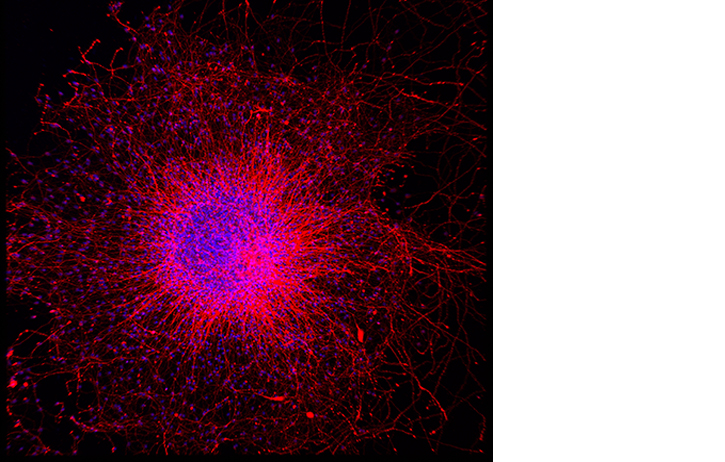
May 2014: Becoming Neurons
Submitted by Louise Collins, Suzanne Crotty and Yvonne Nolan
The birth of new neurons from neural stem cells occurs in the embryo and continues throughout adulthood in discreet regions of the mammalian brain. Neural stem cells group together in ball-like clusters called neurospheres and can be induced to give rise to neurons. Neurospheres are of great interest therapeutically as they could theoretically be used to generate and replace the neurons that are lost in traumatic brain injury and in neurodegenerative diseases such as Parkinson’s disease, Alzheimer’s disease and multiple sclerosis.
Image: This neurosphere was prepared from neural stem cells extracted from an embryonic rat midbrain. After allowing the cells to proliferate and expand for 7 days, they were then imaged with confocal microscopy. Cells differentiated to neurons are labeled red with an antibody to βIII-tubulin. All nuclei are stained blue with bisbenzamide.
“Becoming Neurons” was recently on display in the Jennings Gallery, as part of the “It’s a Beautiful World” Exhibition.
April 2014
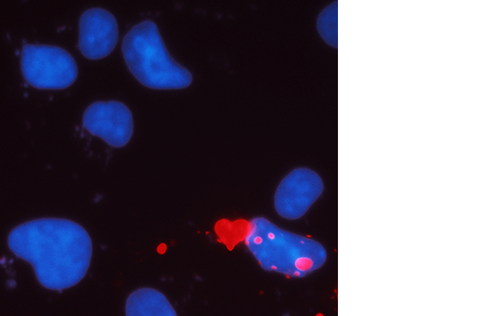
April 2014: Au Coeur de la Cellule
Image submitted by Dr André Toulouse, Department of Anatomy & Neuroscience UCC
Heart-shaped protein aggregates at the periphery of the nucleus of a cell.
Polyglutamine diseases are a group of neurological movement disorders including Huntington’s disease and several spinocerebellar ataxias. We created a cellular model to study abnormal protein production in these diseases and while characterizing the cells under fluorescence microscopy, we observed this heart-shaped protein inclusion at the periphery of a nucleus.
“Au Coeur de la Cellule” was recently on display in the Jennings Gallery, as part of the “It’s a Beautiful World” Exhibition.
March 2014
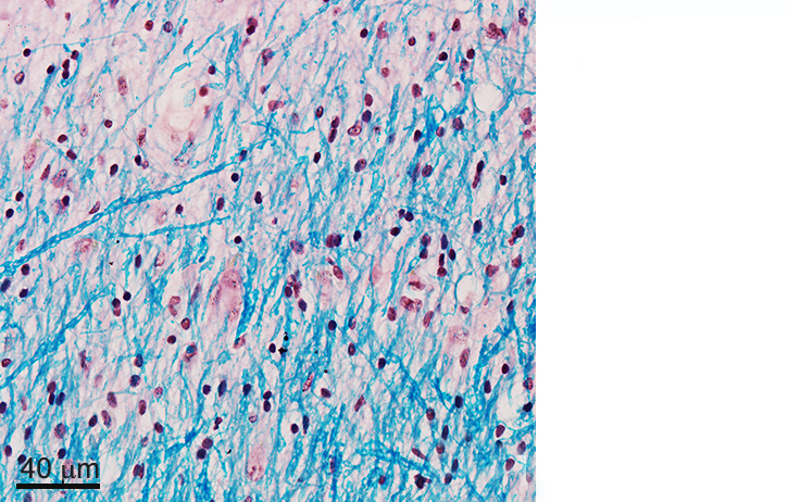
March 2014 Title: “Myelin is getting on my nerves"
This is a post-mortem image collated as part of a HRB-funded summer scholarship awarded to current 4th year Neuroscience BSc student Mr. Richard Magee. Richard undertook this research with Dr. Eric Downer in collaboration with Dr. Yvonne Nolan Department of Anatomy and Neuroscience, UCC.
The image is a light microscopic image of part of the superior frontal gyrus, a region in the frontal lobe of the human brain, of an individual with Primary-Progressive Multiple Sclerosis (MS). Luxol fast blue (LFB) stain has been used to demonstrate the intricate pattern of myelin observed under light microscopy. Using this stain myelin fibres appear blue. LFB has been used in combination with a common hematoxylin and eosin stain. Hematoxylin is a purple dye that stains nuclear material, while eosin is an orange to red dye that stains the cytoplasm. Myelin is a substance rich in lipids and proteins that insulates nerve fibres, allowing efficient transmission of nerve impulses. In MS, an abnormal immune reaction is believed to initiate an attack on the myelin, resulting in its destruction and the appearance of lesions (bare spots and scarred areas along the nerve) throughout the brain. This images shows a region of the human brain that is lesion free, and hence we can observe the extensive network of myelin.
Grant support from the Health Research Board (HRB) is acknowledged. Tissue samples were supplied by the Multiple Sclerosis Society Tissue Bank, funded by the Multiple Sclerosis Society of Great Britain and Northern Ireland, registered charity 207495.
“Myelin is getting on my nerves” image is on display in the Jennings Gallery until 27th March 2014 as part of the “It’s a Beautiful World” Exhibition.
February 2014
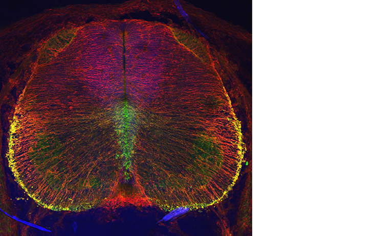
February 2014
Image by Dr Janelle Pakan.
Dr Palkan working in the McDermott lab produced this Confocal image of Rat Spinal cord using the OLYMPUS FV 1000 Confocal microscope.
January 2014
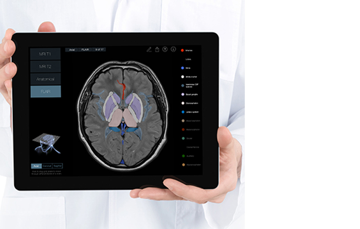
January 2014
Image submitted by Dr Kevin Murphy. This is a promotional image for the Radiology-Head iPod app developed by Dr Murphy.
Dr Murphy is a College Lecturer of Anatomy & Radiology in the Department of Anatomy & Neuroscience, he believes that this app will prove invaluable for clinical reference and patient education. It will be an important resource for medical doctors, healthcare professionals and students preparing for exams. It may also be helpful for patients with neurological diseases and their families in gaining a deeper understanding of their illness.
In the app Radiology-Head, anatomically identical scans of the brain are labelled to allow for comparison of the same anatomical landmarks on different scan types. It is possible to navigate through the head by pulling a 3D plane (Axial, Coronal or Sagittal) through a 3D head to virtually slice through the brain. The 3D Model of the head also allows the user to easily identify the orientation and location of the scan.
Dr Murphy along with former anatomy lecturer Dr Lee Crush, Professor Michael Maher & Dr Owen O'Connor from the UCC Department of Radiology worked closely with 3D4Medical to develop the app. 3D4Medical are world leaders in development of visually appealing medical apps for the iPad and iPhone.
The app Radiology-Head is available to purchase through iTunes.
December 2013
December 2013
Neurosphere Image by Yvonne Nolan, Louise Collins, Suzanne Crotty, Department of Anatomy and Neuroscience has been included in CELL PICTURE SHOW, an online forum hosted by the journal "Cell".
November 2013
November 2013
Image by Dr Denis Barry
October 2013
October 2013
September 2013
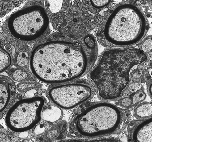
Image by Dr Don O Leary
Transmission electron micrograph of myelinated rat spinal nerves