In This Section
- Home
- Staff Profiles & Phone Book
- About the Department
- Welcome from Head of Department of Anatomy and Neuroscience
- A History of the Department
- A history of the Department; The early years to the 1980s
- A history of the Department; The move from the Windle Building to BSI and WGB
- UCC Professors of Anatomy and Heads of Department
- The development of the UCC HUB
- Current students, recent research graduates and awards
- Useful Links
- Study Anatomy
- Study Neuroscience
- Research
- Neural circuitry underlying Neuropsychiatric and Neurological Disorders 2026
- Neurogastroenterology 2026
- Developmental Neuroscience and Regeneration 2026
- Neurodegeneration 2026
- Neuroinflammation 2026
- Neuroprotection and Therapeutics 2026
- Neuroproteomics and Molecular Psychiatry 2026
- Anatomy Education Research 2026
- Research Facilities 2026
- Postgraduate Research Programmes 2026
- UCC Anatomical Donations
- Biosciences Imaging Centre
- BSc Medical and Health Sciences
- News & Events
- News Archive 2024
- News Archive 2023
- News Archive 2022
- News Archive 2021
- News Archive 2020
- News Archive 2019
- News Archive 2018
- News archive 2017
- News Archive 2016
- News Archive2015
- News Archive 2014
- News Archive 2013
- News Archive 2012
- News Archive 2011
- BRAIN AWARENESS WEEK 2023
- Department Events and Conferences
- Seminar series 2019_2020
- photo galleries
- Narrowing the void Conference 2023
- Photos of BSc Medical and Health Sciences Mentoring launch 2022
- International Women's Day 2023
- 2023 BRIGHT FUTURES - Celebrating our researchers
- 2023 UCC Futures - Future Ageing & Brain Sciences
- Recent Graduations July 2023
- Anatomy and Neuroscience Top 100 Anatomy Physiology 2023
- BRAIN AWARENESS WEEK 2023 FUN AND GAMES EVENT
- Medical and Health Sciences First year class 2023
- 2023 Brain Awareness week Scientific discussion photo gallery
- World Anatomy Day 2023
- BSc MHS MENTORING PROGRAMME 2023
- BSc Medical and Health Sciences Graduation 2023
- BSc Neuroscience Graduation Photo Gallery 2023
- Dr Kathy Quane Nov 2023
- THANKSGIVING PHOTOS 2012
- Photo Gallery: Society of Translational Medicine Careers Fair 2023
- Photo Gallery:2023 TRAIN AWARDS
- Photo Gallery:2024 Creative Week St Joseph's NS
- Photo Gallery: Department of Anatomy and Neuroscience Thanksgiving Service 2024
- Photo Gallery: Professor Aideen Sullivan farewell party
- Photo Gallery: Irish Pain Society Annual Scientific Meeting Cork 2023
- Photo Gallery: 2024 Medical and Health Sciences Graduation
- Photo Gallery: Medical and Health Sciences Meet and Greet 2024
- Photo Gallery: 2024 BSC NEUROSCIENCE Graduation
- Photo Gallery: 2025 INTERNATIONAL WOMEN'S DAY
- Photo Gallery: 2025 BSc Neuroscience class and staff
- Photo Gallery: 2025 BRAIN CONNECTIONS
- BSc Neuroscience Graduation Photo Gallery 2025
- World Anatomy Day 2025
- UCC Learning and Teaching Showcase 2025
- MSc Human Anatomy Graduation Photo Gallery 2025
- Narrowing the Void Conference 2023
- Department of Anatomy and Neuroscience Contact Us
October 2015
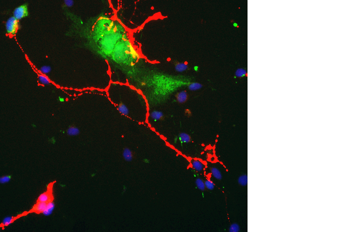
October 2015: Embryonic ventral midbrain dopaminergic neuron (red) synpases on a transfected non-dopaminergic neuron (green) expressing green fluorescent protein.
Submitted by: Shane V. Hegarty Post-Doctoral Research Fellow, Department of Anatomy and Neuroscience.
Dr Shane Hegarty is a BSc and PhD graduate from the Department of Anatomy and Neuroscience, UCC, who recently began an Irish Research Council Post-Doctoral Research Fellowship under the supervision of Dr Aideen Sullivan and Dr Gerard O’Keeffe. Their research investigates the role of BMP-Smad signalling in ventral midbrain dopaminergic neuron development.
Transfection allows the introduction of specific sequences of nucleic acids, e.g. DNA or RNA, into a desired cell type, and is generally employed to alter the expression levels of a protein of interest. Transfection of primary neurons enables the investigation of the functional roles of various target genes in neuronal development and survival. These transfected neurons can be visualised through the ectopic expression of fluorescent proteins, such as green fluorescent protein (GFP).
The image depicts an embryonic ventral midbrain dopaminergic neuron (red) which has formed synapses on a transfected non-dopaminergic neuron (green), expressing green fluorescent protein, in a culture of the embryonic ventral midbrain following four days of differentiation. Synapses are the point of electrochemical communication between neurons. Embryonic day 14 ventral midbrain cells were transfected via electroporation with specific plasmid(s), and subsequently differentiated using serum proteins. A transfected glial cell (green) also appears to be in contact with the processes of another dopaminergic neuron (red). Cellular nuclei were stained with bisbenzamide (blue). The image was taken using an inverted fluorescence microscope at 200x magnification.
This image was recently exhibited as part of the BRAINTALK Parkinson’s Community Meeting and Art Exhibition, held in the Glucksman Gallery University College Cork. This event was organised by Dr Shane Hegarty and Professor Aideen Sullivan of the Department of Anatomy and Neuroscience, UCC, as part of the BRAINTALK project (www.ucc.ie/en/braintalk).
The purpose of the meeting was to bring together People with Parkinson's, Parkinson's researchers and clinicians, to create an interconnected Parkinson's Community in Ireland. Over 200 participants, the majority of whom were People with Parkinson’s and their carers, gathered in the Glucksman Gallery for a variety of presentations by Parkinson’s advocacy groups, People with Parkinson’s, therapists, neurologists and neuroscientists.
‘Parkinson’s Community’ art exhibition was launched after the meeting. This included paintings made by People with Parkinson’s in an ‘Exploring Parkinson’s with Art’ workshop , as well as photomicrographs showing the ongoing scientific work by Parkinson’s researchers in the Department of Anatomy and Neuroscience, UCC.
For full details see BRAINTALK website http://www.ucc.ie/en/braintalk/.
Department of Anatomy and Neuroscience
Anatamaíocht agus Néareolaíocht
Contact us
Room 2.33, 2nd Floor, Western Gateway Building, University College, Cork, Ireland