- Home
- Staff Profiles & Phone Book
- About the Department
- Study Anatomy
- Study Neuroscience
- Research
- UCC Anatomical Donations
- Biosciences Imaging Centre
- BSc Medical and Health Sciences
- News & Events
- Contact Us
SPOTLIGHT EVENTS
Microscopy image chosen for CELL PICTURE SHOW
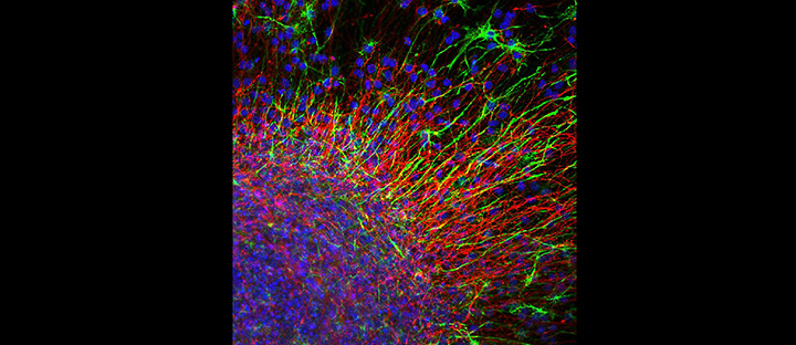
The image by Yvonne Nolan, Louise Collins, Suzanne Crotty, Department of Anatomy and Neuroscience, UCC can be viewed at
When cultured with growth factors in vitro, neural stem cells can generate neurons (red), as well as the cells that support them— astrocytes (green) and oligodendrocytes. In culture, neural stem cells group together in ball-like clusters, called neurospheres (bottom left). Neurospheres are of great therapeutic interest because they have the potential to regenerate and replace neurons lost in traumatic brain injury and neurodegenerative diseases, such as Parkinson's disease, Alzheimer's disease, and multiple sclerosis.
Image: This neurosphere (bottom left) was prepared from neural stem cells extracted from an embryonic rat midbrain. After allowing the cells to proliferate and expand for 7 days, they were imaged with confocal microscopy. Cells differentiated to neurons are labeled red with an antibody to βIII-tubulin, whereas astrocytes are labeled green with an antibody to glial fibrillary acidic protein (GFAP), an intermediate filament specific to astrocytes. All nuclei are stained blue with bisbenzamide.