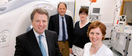
Cork doctors have shown that the radiation exposure of patients with Crohn’s disease referred for a CT scan can be significantly reduced while retaining diagnostic accuracy.
CT (computed tomography) is the preferred method for assessing the complications of active Crohn’s disease. However, the downside to traditional CT scans is radiation, which might become hazardous particularly if repeated scanning is required. While imaging strategies that do not involve radiation, such as MRI (magnetic resonance imaging), are being explored, the recommended imaging in many situations remains CT scanning.
Clinician-scientists in Cork have collaborated to devise a new CT scanning protocol, using recently developed computer software, for imaging of the abdomen and pelvis in patients with Crohn’s disease and this has resulted in the lowest doses of diagnostic radiation ever reported in this clinical setting, whilst maintaining diagnostic accuracy. The research was carried out at Cork University Hospital and the Science Foundation Ireland-funded Alimentary Pharmabiotic Centre at University College Cork and has been reported in the journal Clinical Gastroenterology & Hepatology.
The research group in Cork first identified a potential problem with diagnostic radiation exposure and previously reported that patients with inflammatory bowel disease (IBD), particularly those with Crohn’s disease, but also including other gastrointestinal disorders, may be exposed to high levels of radiation—mostly from abdominal/pelvic computed tomography (CT) scans. The new CT protocol was compared with conventional CT and was found to reduce radiation exposure by over 70% from that of a conventional abdominal/pelvic CT scan without loss of diagnostic accuracy.
Prof. Michael Maher who led the research said that “the most important result was that our direct comparative trial of low vs conventional dose CT scanning showed we could retain diagnostic accuracy whilst reducing radiation exposure”. He also said that “the research is continuing and we anticipate further improvements in the quality of the imaging as we refine the protocol with the latest computer software technology.” He also emphasized that currently the results apply only to Crohn’s disease and cannot be extrapolated to other conditions at this stage.
“This is good news for patients with Crohn’s disease, particularly those who need repeat CT scanning for management of the complications of their illness. This research is an important step forward and is already changing clinical practice,” said Prof. Fergus Shanahan, one of the authors of the scientific article.
Crohn’s disease is a lifelong, relapsing and remitting disorder which may affect any part of the bowel. Patients often require surgical operations as well as anti-inflammatory and immunosuppressive drug treatment.
The research is published in the journal clinical Gastroenterology and Heaptology; authors Craig O, O'Neill S, O'Neill F, McLaughlin P, McGarrigle A, McWilliams S, O'Connor O, Desmond A, Walsh EK, Ryan M, Maher M, Shanahan F. “Diagnostic Accuracy of Computed Tomography Using Lower Doses of Radiation for Patients with Crohn's Disease” Clin Gastroenterol Hepatol. 2012 [Epub ahead of print] PMID: 2246999
« Previous Item
Next Item »
« Back to 2012 Press Releases
